


 https://orcid.org/0009-0000-2198-4836

Abstract
Abdominal ectopic pregnancy is a rare and potentially life-threatening condition requiring timely diagnosis and coordinated surgical care. We report the case of a 28-year-old woman, gravida 2, abortus 1, para 0 (G2A1P0), at 17 weeks’ gestation, identified during routine antenatal care with suspected abdominal pregnancy. Ultrasound findings prompted further evaluation with magnetic resonance imaging, which clarified the implantation site and confirmed the diagnosis. On admission, she was hemodynamically stable, with mild anaemia and elevated β-hCG levels. Following multidisciplinary planning and preparation for possible massive transfusion, elective laparotomy revealed the gestation within the left rudimentary uterine horn beneath the meso-sigmoid colon. The fetus and gestational structures were removed, and mesocolon repair was completed without complication. The patient recovered uneventfully and was discharged on postoperative day 10. This case underscores the importance of advanced imaging, meticulous surgical planning, and multidisciplinary collaboration in managing abdominal ectopic pregnancy.
Introduction
Ectopic pregnancy is a serious condition in which implantation occurs outside the uterine cavity. While most are tubal, abdominal ectopic pregnancies are rare, accounting for ~1% of cases, and are associated with high maternal morbidity and mortality [1–4]. Advances in imaging such as ultrasound and magnetic resonance imaging (MRI) have improved diagnostic accuracy, but management remains challenging.
Case presentation
A 28-year-old woman, gravida 2, abortus 1, para 0 (G2A1P0), at 17 weeks’ gestation, presented to the National Hospital for routine antenatal care. Obstetric ultrasound revealed an abdominal ectopic pregnancy (Fig. 1). She was admitted for further evaluation. On admission, physical examination showed a blood pressure of 127/79 mmHg, normal pulse rate consistent with obstetric condition, body temperature of 37°C, and normal level of consciousness. Abdominal examination revealed a distended abdomen without tenderness on palpation.
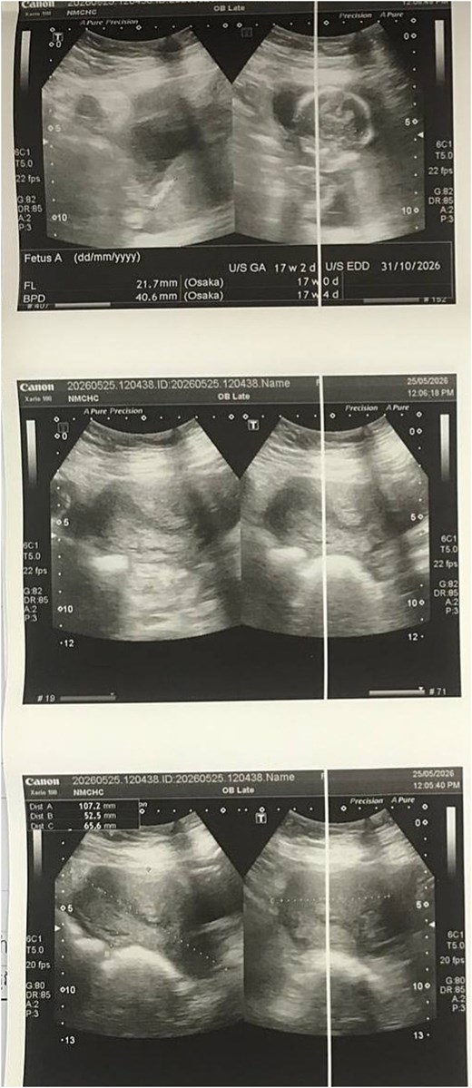
Ultrasound demonstrating an abdominal ectopic pregnancy.
Laboratory testing demonstrated a serum β-hCG level of 43 262 mIU/ml, with haematology showing mild anaemia (haemoglobin 10.7 g/dl, haematocrit 34.4%) and elevated red cell distribution width (RDW-CV 14.2%). White blood cell count (9.3 × 10⁹/L), platelets (261 × 109/L), and differential counts were within normal limits. Serology was negative for syphilis, HIV, and hepatitis B (Table 1). Urinalysis was unremarkable. Her blood group was B Rh(D) positive. MRI was performed to clarify the implantation site of the ectopic pregnancy and confirmed an abdominal ectopic gestation (Fig. 2).
Laboratory results
| Test | Result | Unit | Reference range | Flag |
|---|---|---|---|---|
| WBC | 9.3 | ×109/L | 4.0–10.0 | Normal |
| RBC | 3.95 | ×1012/L | 3.8–5.4 | Normal |
| Haemoglobin | 10.7 | g/dl | 11–15.5 | Low |
| Haematocrits | 34.4 | % | 35–48 | Low |
| MCV | 87.1 | fl | 80–100 | Normal |
| MCH | 27.1 | pg | 25–33 | Normal |
| MCHC | 31.1 | g/dl | 28–36 | Normal |
| Platelets | 261 | ×109/L | 150–500 | Normal |
| RDW-CV | 14.2 | % | 11.5–14.0 | High |
| Neutrophils | 6.5 | ×109/L | 2–7.5 | Normal |
| Lymphocytes | 2.2 | ×109/L | 1–4 | Normal |
| Monocytes | 0.4 | ×109/L | 0.2–1.0 | Normal |
| Eosinophils | 0.2 | ×109/L | 0.02–0.5 | Normal |
| Basophils | 0.0 | ×109/L | 0–0.1 | Normal |
| Beta HCG | 43 262 | mIU/ml | 0–5 | High |
| Blood group | B Rh(D) positive | |||
| RPR (syphilis) | Negative | |||
| HIV 1/2 Antibody | Negative | |||
| HBsAg (Hepatitis B) | Negative | |||
| Urine protein | Negative | |||
| Urine glucose | Negative |
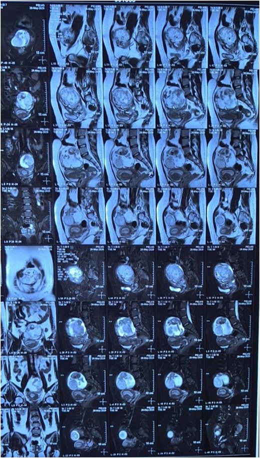
MRI confirming abdominal ectopic pregnancy.
Given the anticipated complexity of surgery, full preparation was made for the possible need of massive blood transfusion. Following multidisciplinary consultation involving gynaecology–obstetrics, general surgery, and anaesthesia teams, elective laparotomy under general anaesthesia was undertaken. Intraoperatively, the ectopic gestation was identified within the left rudimentary uterine horn, located beneath the meso-sigmoid colon (Fig. 3). Dissection was performed to isolate the ectopic pregnancy, with meticulous dissection of the meso-sigmoid colon. The mass included the placenta, gestational sac, and fetus. A dead fetus weighing 180 g was extracted from the gestational sac (Fig. 4). No colonic injury occurred, and the mesocolon was repaired (Fig. 5). The abdominal cavity was irrigated, and a drain was placed in the pouch of Douglas.
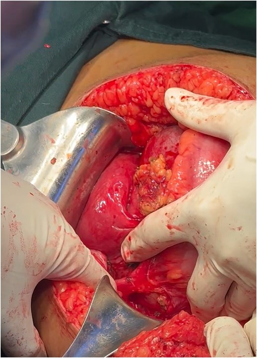
Intraoperative findings of abdominal ectopic pregnancy.
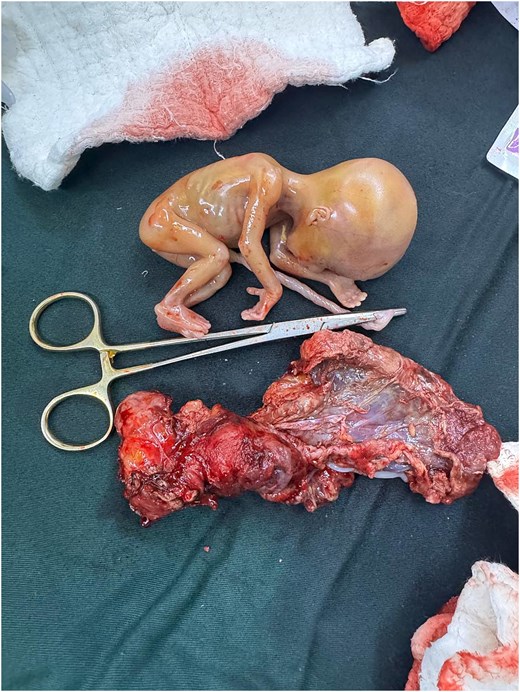
Extracted dead foetus from the gestational sac.
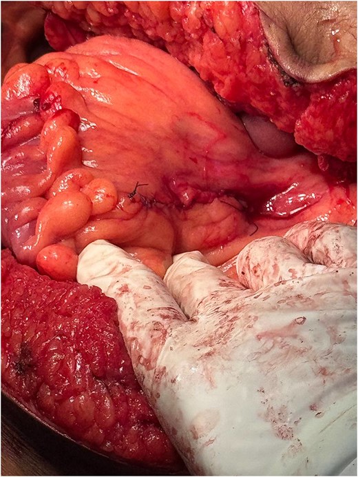
Mesocolon repair following ectopic mass dissection.
The patient tolerated the procedure well. Postoperatively, serum β-hCG levels declined appropriately. Her recovery was uneventful, with good wound healing, and she was discharged on postoperative day 10 in stable condition.
Discussion
Abdominal ectopic pregnancy is rare and often presents later in gestation compared to tubal ectopic pregnancies. Diagnosis is challenging because symptoms are non-specific and may mimic other intra-abdominal pathologies. Clinical suspicion should be raised in patients with atypical pain, inconclusive imaging, or abnormal gestational findings [5]. Ultrasound remains the first-line modality, but its sensitivity decreases with unusual implantation sites. MRI provides superior localization and delineation of anatomical relationships, making it invaluable for preoperative planning [3].
Reported implantation sites are diverse, reflecting the complexity of diagnosis and management. Documented locations include the posterior cul-de-sac [6], sigmoid mesocolon (as in the present case), uterine horn [7], omentum, retroperitoneum [8], broad ligament, and even visceral organs such as the liver, spleen, and bowel surfaces [3]. These varied sites highlight the importance of advanced imaging and intraoperative vigilance.
Management decisions depend on gestational age, implantation site, patient stability, and available expertise.
Early gestations with non-viable pregnancies may be candidates for expectant management [8].
Mid-trimester or viable pregnancies typically require surgical intervention due to risk of haemorrhage and maternal morbidity [9].
Multidisciplinary planning is essential, involving obstetricians, general surgeons, anaesthesiologists, and radiologists to anticipate intraoperative challenges.
In this case, the decision for elective open surgery was guided by gestational age (17 weeks), anatomical complexity (implantation beneath the sigmoid mesocolon), and the need for secure haemostasis.
Surgical excision remains the cornerstone of treatment.
Laparotomy is preferred in advanced gestations or complex anatomical sites [6, 7, 9].
Laparoscopy may be feasible in selected early cases, offering reduced morbidity [6].
Haemostasis is critical, as implantation sites are often highly vascular. Surgeons must be prepared for potential massive haemorrhage, and blood products should be available.
Postoperative monitoring of β-hCG levels ensures complete removal of trophoblastic tissue.
This case demonstrates the importance of individualized management, advanced imaging for diagnosis, and multidisciplinary collaboration. Literature reports highlight diverse implantation sites and management strategies, underscoring the need for clinician awareness [1, 7, 9, 10].
Conclusion
Abdominal ectopic pregnancy is a rare but life-threatening condition. Early diagnosis with ultrasound and MRI, combined with timely surgical intervention, is critical for favourable outcomes. Clinicians should maintain vigilance for non-tubal implantation sites, especially in atypical presentations.
Acknowledgements
The author wishes to express sincere gratitude to the Director of the National Maternal and Child Health Centre (NMCHC), Phnom Penh, Cambodia, for institutional support and guidance. Special thanks are extended to the surgical team and anaesthesia team for their dedicated clinical care and collaboration, which made this study possible.
Author contributions
Chansokha Soeur (Conceptualization [lead], Writing—review & editing [lead]).
Conflicts of interest
The authors declare no conflicts of interest.
Funding
None declared.
Data availability
All data supporting the findings of this case report are included in the manuscript. Additional anonymized information may be made available from the corresponding author upon reasonable request and with institutional approval.
Ethical approval
This study received ethical approval from the Institutional Review Board of the National Maternal and Child Health Centre (NMCHC), Phnom Penh, Cambodia. All procedures were conducted in accordance with institutional guidelines and the ethical principles of the Declaration of Helsinki.


