


 https://orcid.org/0000-0003-0001-9070

Abstract
Peritoneal dialysis catheters are an excellent and highly effective treatment for end-stage renal failure. This procedure is relatively easy and straightforward, but as with any other medical procedure, it can lead to complications, which include infection, hematoma, leakage, and peritonitis. However, in a few selected cases, the catheter can perforate the bowel, and in even rarer cases, it can migrate through it.
We report the case of an 18-year-old who needed renal replacement therapy through a Tenckhoff catheter. Two years after its placement, he suddenly noticed that the tip of the catheter protruded through his anus. Migration of the catheter was diagnosed, and during surgery, a fistulous tract from the small bowel to the rectum was found. After surgery, the patient recovered successfully and is on close follow-up.
Introduction
Peritoneal dialysis catheters are an excellent option for end-stage renal failure [1, 2]. This catheter is relatively safe and can help many patients [2, 3]. Nonetheless, as with any medical procedure, nothing is without risk. Infection, hematoma, leakage, and peritonitis are the most common [1, 2]. However, in a few selected cases, the catheter can perforate the bowel, and in even rarer cases, it can migrate through it [1]. Various factors, including omental attachment, bowel contractions, peritoneal adhesions, and the volume of dialysis fluid, can aid in the peritoneal catheter migration [1, 2].
This extremely rare complication, with a few cases reported in the English literature, can cause severe morbidity and leave the catheter useless if left untreated [2]. Prompt intervention and high clinical awareness are needed in these patients [2, 3].
We present the case of an 18-year-old male with stage V chronic kidney disease who had peritoneal dialysis through a Tenckhoff catheter. He noticed that the catheter tip protruded from his anus. After further evaluation, catheter migration was diagnosed and treated.
Case report
Patient is an 18-year-old with a past medical history of systemic lupus erythematosus and diffuse lupus nephritis, which led to stage V chronic kidney disease that needed peritoneal dialysis as a renal replacement therapy through a Tenckhoff catheter.
Throughout this time, he remained completely asymptomatic and had dialysis without complications; nevertheless, 2 years after its placement, one day, he noticed that the tip of the catheter protruded through his anus, therefore, he presented to the emergency room.
On clinical evaluation, the patient was completely asymptomatic, and on a rectal exam, we confirmed the presence of the Tenckhoff catheter tip in the rectum (Fig. 1a). As a result, complementary exams were required, and a contrast-enhanced abdominal computed tomography (CT) confirmed the bowel perforation and transrectal migration into the rectum (Fig. 1b and c). Other exams, including complete blood count and C-reactive protein, were normal. With these findings, the patient was admitted to the hospital, a central venous catheter was placed for hemodialysis, and surgery was decided with his parents.
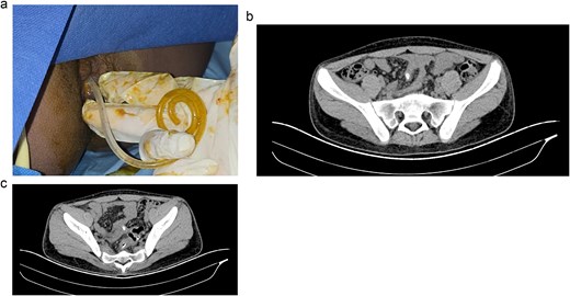
(a) Tenckhoff catheter is seen through the anus. (b) CT, the catheter is seen in the intestinal lumen. (c) CT, the catheter is seen in the rectum.
On laparoscopy, we discovered multiple adhesions, which were bluntly dissected; the catheter created a fistulous tract into the small bowel, which was located in the ileum, 20 cm away from the ileocecal valve. At this point, the catheter was fused with the small bowel and partially obstructed its lumen. Afterward, it exited the bowel and perforated the upper rectum. Then it exited through the anus (Fig. 2a–c).
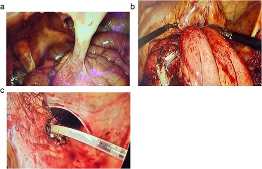
(a) The catheter is seen in the small bowel with a fistulous tract. (b) Multiple adhesions are seen in the bowel with the catheter. (c) The catheter is seen in the rectum.
With these findings, the entire fistula was removed. We resected the terminal ileum with a mechanical stapler (Echelon, NJ, Johnson & Johnson), removed the catheter, and then identified the site of the rectal perforation. The catheter was then pulled out through the anus, and the rectum was divided at the site of perforation. To restore bowel continuity, a recto-rectal anastomosis was completed, and a protective ileostomy was completed without complications. A drain was left in place, and the procedure was finalized without complications.
The patient's postoperative course was uneventful. He remained in the surgical ward without complications, and a peripheral central line was placed for hemodialysis. He was discharged after 3 days, and 6 months later, he is on close follow-ups for intestinal ostomy reversal surgery.
Discussion
Peritoneal dialysis is an effective method to provide a safe and accessible treatment for renal replacement therapy to many patients with impaired renal function [1, 2]. Moreover, since the global burden of kidney disease continues to increase, so does the need for a cost-effective treatment, such as peritoneal dialysis [2]. Since our patient had renal disease and we are in a developing country where costs are a constraint, peritoneal dialysis was the most cost-effective treatment.
Nonetheless, as with almost every surgical and invasive procedure, none is without risk, and complications from peritoneal dialysis can appear in up to 10% of patients [2, 3]. Most complications occur early during catheter insertion and are usually due to a malfunction, placement, or infection [1, 4]. Perforation and delayed perforation are infrequent complications that occur months (1.6–48) after surgery, most commonly when the catheter has not been used for quite some time [2, 4].
This uncommon event has been poorly explained in the literature; however, it's believed that delayed perforation most likely occurs when the catheter is in the peritoneal cavity and not in use [3–5]. Without liquids to bathe the bowel loops and the catheter, the catheter does not have a barrier to prevent adhesions to the bowel wall [2, 4]. As the catheter attaches to the bowel, it exerts continuous pressure, leading to ischemia, erosion, laceration, and perforation [6]. Other risk factors include diverticular disease or any other gastrointestinal pathology, such as amyloidosis [3, 7].
Since our patient had limited resources, this may have limited his access to healthcare, thereby delaying his dialysis and contributing to catheter migration.
Delayed perforations have been described in multiple viscera, including the rectum, the bladder, the urethra, and the vagina [3, 4]. When a delayed perforation occurs, symptoms are heterogeneous and range from asymptomatic patients to those resembling peritonitis, diarrhea, or protrusion of the catheter from the anus or intestinal discharge [3, 8], as it happened to our patient.
Treatment is straightforward: the catheter must be removed and bowel repair completed [2]. A conservative approach could be performed; however, if there are signs of sepsis or peritonitis, surgical intervention is necessary via a laparoscopic or conventional approach [2, 3, 9].
In our case, we had a complex fistula in which two segments of the bowel were compromised. As this patient had multiple comorbidities, and due to the risk of leaks, an ostomy was performed; afterward, he overcame this event in one close follow-up.
Conclusion
Peritoneal dialysis catheters are extremely useful for patients with kidney disease; however, like any chronic treatment, they require ongoing monitoring. This case demonstrates that every patient under our care requires close monitoring, despite all the challenges faced by the healthcare system. Patient education prior to any treatment is vital in order to diagnose and prevent complications.
Conflicts of interest
None declared.
Funding
None declared.


