


 https://orcid.org/0009-0001-6978-9703

Abstract
Eosinophilic solid and cystic renal cell carcinoma (ESCRCC) is a rare entity recently included in the world health organization classification of kidney tumors. Due to its indolent behavior and insufficient characterization, it is often misdiagnosed. This report describes the case of a 28-year-old Ghanaian female who presented with a 1-year history of a gradually enlarging, initially painless right flank mass detected on self-examination, without any hematuria. A contrast-enhanced abdomino-pelvic computerized tomography scan demonstrated a well-defined, heterogeneously enhancing mixed solid and cystic lesion measuring 9.9 × 7.3 × 9.0 cm arising from the interpolar region of the right kidney. She had open radical right nephrectomy. Post-operative pathological immunohistochemical staining diagnosed ESCRCC. She is currently doing well after 8 months with no concern for metastasis. Initial misdiagnosis could negatively impact patient outcome and therefore underscores the need for early and definitive diagnosis of ESCRCC.
Introduction
Renal cell carcinoma (RCC) accounts for a small but considerable number of adult malignancies, with an expanding spectrum of histologic subtypes distinguished by distinctive morphologic, immunophenotypic, and molecular characteristics [1]. RCC represents ~90% of all renal malignancies and is increasingly identified incidentally on routine imaging [2]. The World Health Organization’s (WHO) 2022 classification of kidney tumors highlights diagnostic advancements with inclusion of three new entities. Among these, eosinophilic solid and cystic renal cell carcinoma (ESCRCC) has emerged as a rare subtype [3]. This tumor was first described by Trpkov and colleagues in 2016 after observing a unique form of RCC with eosinophilic cytoplasm and varying degrees of solid and cystic components [4]. Although the vast majority of ESCRCC occur sporadically, some cases are associated with tuberous sclerosis complex (TSC) genes 1 and 2 mutation (10%), predominantly occurring among females [5]. Due to its indolent behavior and insufficient characterization, there may be an overlap with other oncologic neoplasms that commonly leads to misdiagnoses. Pathological assessment remains the most reliable diagnostic method [4]. We report a 28-year-old Ghanaian female initially diagnosed twice with renal oncocytoma before a final diagnosis of ESCRCC.
Case report
A 28-year-old woman presented with a 1-year history of a gradually enlarging, initially painless right flank mass detected on self-examination. Associated symptoms included dizziness, fatigue, back pain without hematuria, fever, or weight loss. She used non-steroidal anti-inflammatories for analgesia and had a 7 years history of second-hand tobacco smoke exposure. There was no family history of renal tumors or hereditary cancer syndromes. Physical examination revealed a mildly tender, firm right flank mass measuring ~13 × 11 cm extending into the lumbar region.
Contrast-enhanced abdomino-pelvic computerized tomography (CT) scan demonstrated a well-defined, heterogeneously enhancing mixed solid and cystic lesion measuring 9.9 × 7.3 × 9.0 cm arising from the interpolar region of the right kidney, with intralesional calcifications and radiologic features consistent with a Bosniak class IV (Fig. 1) [6].
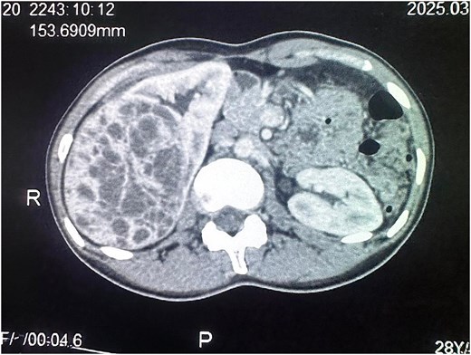
Axial cut of contrast-enhanced CT scan of the abdomen at the level of the kidneys. A heterogeneously enhancing mixed solid and cystic lesion measuring 9.9 × 7.3 × 9.0 cm arising from the interpolar region of the right kidney is visible.
Patient subsequently underwent an open radical right nephrectomy, which revealed a large right renal mass with no gross involvement of the renal vein or inferior vena cava and no obvious regional lymphadenopathy (Fig. 2).
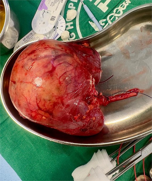
Appearance of a large renal mass on macroscopic pathological examination. The specimen weighed 554 grams and measured 12 × 7.8 × 7 cm with a 5.5 cm long ureter attached. The tumor was well circumscribed with a grossly intact capsule.
Histopathological examination (HPE) showed a tumor weighing 554 g and measuring 12 × 7.8 × 7 cm with a 5.5 cm long ureter attached. The tumor involved the upper and middle pole of the right kidney, without lymph node involvement. It was well circumscribed with a pale brown cut surface containing multiple cystic areas and an intact capsule. The tumor was macroscopically and microscopically limited to the kidney with no lymphovascular invasion or positive margins. It demonstrated solid, tubular, micro, and macrocystic patterns. The lesional cells were relatively uniform with abundant dense eosinophilic cytoplasm with rare mitoses. Initial diagnosis was renal oncocytoma, with no rhabdoid, sarcomatous or necrotic features. Immunohistochemistry (IHC) with CD117 and Cytokeratin-7 (CK7) stains were negative with no nuclear or cytoplasmic staining and a diagnosis of eosinophilic cell cytoplasm made. Further IHC with AMACRACEMASE/P5045 (alpha methyl acyl CoA racemase) (AMACR) stain was positive (Table 1), leading to a final diagnosis of ESCRCC.
Immunohistochemical results.
| Stains | Results | Details |
|---|---|---|
| CD117 | Negative | No nuclear or cytoplasmic staining |
| CK7 | Negative | No nuclear or cytoplasmic staining |
| AMACR | Positive | Diffuse, moderate nuclear or cytoplasmic staining |
At 8 month follow-up, she is clinically well without any evidence of metastasis.
Discussion
ESCRCC is a rare renal neoplasm officially included in the 2022 WHO classification of kidney tumors [3, 5, 7]. Historically termed unclassified or TSC-associated RCC, it accounts for ~0.2% of renal tumors and was only recently defined as a separate morpho-molecular entity [3, 8]. Less than 70 cases have been reported worldwide with majority presenting as localized lesions and few demonstrating malignant potential [8, 9].
This case exemplifies the diagnostic challenges encountered by this 28-year-old Ghanaian woman who initially received two diagnoses of renal oncocytoma before a definitive ESCRCC diagnosis following a third pathological review and IHC staining over seven months. Such delays illustrate the difficulty in recognizing this entity, especially in West Africa where reporting and awareness remain limited.
ESCRCC predominantly affects middle aged women with an average age of 57 years but a range of 14–79 years [5]. Cases have also been reported in men and children, including an 8-year-old boy [10]. A noteworthy case in a 19-year-old South African male with typical ESCRCC features presented with an additional focal melanin pigment [11]. Our patient was younger than the average reported age and fits the female predilection, with the male to female ratio estimated between 1:1.2 and 1:1.7 [7]. The tumor in this case was 12 cm in its largest diameter, exceeding the commonly recorded range of 3.4–4.2 cm [9]. However, some isolated case reports identified similar large tumors sizes [12, 13]. Unlike the typical asymptomatic indolent course [8], our patient had mild right flank pains and an abdominal mass.
The histopathological and IHC features observed in this case corresponds closely with established ESCRCC features. Morphologically, ESCRCC exhibits solid and cystic growth patterns with hobnail-cell lined cysts and solid areas containing cells abundant in eosinophilic cytoplasm [5]. Definitive diagnosis hinges on a characteristic immunoprofile featuring CK20 positivity alongside CK7 negativity [3]. In this case, IHC with CD117, CK7, and AMACR enabled exclusion of oncocytoma which is typically CD117 and CK7 positive, and confirmed ESCRCC.
Surgery remains the optimal therapeutic approach and may involve partial or radical nephrectomy. Nephron-sparing surgeries are considered in localized tumors ˂7 cm [10]. Given the tumor size of ~13 × 11 cm, patient had a radical right nephrectomy which was appropriate. Postoperatively, renal function remained normal with no concern for metastasis. In metastatic disease, targeted therapy and immunotherapy have shown success. Palsgrove and colleagues reported complete response with everolimus, an mTOR inhibitor, in a patient with hepatic metastases from ESCRCC [14].
Some limitations observed highlight the challenges in diagnosing rare malignancies in resource constraint settings. Multiple HPE tests were required because of initial misclassification and limited access to specific IHC stains. We relied on the negative CK-7 without confirming CK-20 positivity. Additionally, genetic testing for TSC mutations, more accessible in high resource settings was unavailable. These barriers complicate adherence to the molecular-focused WHO diagnostic criteria and contribute to underreporting in West Africa [3, 15].
In conclusion, this case likely represents one of the few cases seen in West Africa. Although indolent with favorable prognosis, ESCRCC retains malignant potential, highlighting the need for early definitive diagnosis. Pathologists must maintain a high index of suspicion for ESCRCC when evaluating large solid and cystic eosinophilic renal masses. Consistent application of CK20+/CK7- immunoprofile is vital for adequate differentiation and prompt management considering the malignant potential.
Conflicts of interest
The authors declare no conflict of interest.
Funding
No funding was received for this report.


