


Abstract
Benign peripheral nerve sheath tumors are growths that develop in the protective sheath that surround peripheral nerves. In rare circumstances, they can cause mass effect on surrounding structures intra-abdominally. We report an otherwise healthy 62-year-old female who presented for constipation and recurrent bouts of diverticulitis. The patient had known about her nerve sheath tumor but did not want it removed. The initial plan was to perform a robotic Sigmoidectomy; however, this was not feasible given the size of the tumor intra-operatively. The procedure was subsequently aborted. A multi-disciplinary team of vascular surgery, neurosurgery, and colorectal surgery then took the patient to the operating room for an open resection of the schwannoma and concurrent sigmoidectomy. There have only been several staged attempts described in the literature, but none that describe simultaneous resection of both the nerve sheath tumor and the sigmoid colon.
Introduction
Benign peripheral nerve sheath tumors (bPNST) are rare lesions of ectodermal origin that encompass peripheral nerve structures. They can present in a multitude of ways, ranging from asymptomatic to debilitating. Of the bPNSTs, the most common type is the schwannoma, with an incidence rate of up to 75.3% of bPNSTs [1]. These specific tumors may occur sporadically but are often associated with familial syndromes such as Carney’s complex or neurofibromatosis type II [2].
Schwannomas are tumors that undergo degenerative change over time. They have a smooth nodular outline and have a tan or yellow surface. On immunohistochemistry, they show strong expression of S100 protein and abundant pericellular type IV collagen [3] They typically affect the small peripheral nerves in the head and neck, along with flexor surfaces of extremities [3]. Intra-abdominal schwannomas are a rare entity and can cause symptoms secondary to mass effect. In these cases, the schwannoma is resected en bloc [4]. They should be excised with clear margins to prevent recurrence. Several case reports have been published regarding this phenomenon; however, there is no literature that describes schwannoma resection with concurrent diverticulitis.
In patients with diverticulitis, the timing of surgery is constantly changing. Usually, surgery is offered 6–8 weeks after the most recent flare [5]. The operative technique is a sigmoid colectomy, which can be done minimally invasively or open. The operative steps of a sigmoidectomy involve identifying and preserving critical structures, mobilization of the sigmoid colon, division of the inferior mesenteric artery, division of the sigmoid colon with a linear stapler, and the creation of a tension-free anastomosis with a circular stapler [6].
Case report
A 62-year-old female presented to the office for multiple bouts of diverticulitis. Cross-sectional imaging from her most recent bout of diverticulitis showed a peripheral nerve sheath tumor located on the right spinal column that measured 56 × 72 mm (Fig. 1), abutting the iliac vessels, which at the time was not in proximity of the sigmoid colon. The patient had known about this tumor for quite some time. She had met with a neurosurgeon, but given its benign features and her lack of symptoms, no further intervention was offered.
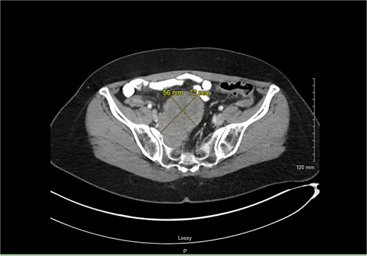
Axial imaging shows the schwannoma with greatest measurements of 7.2 × 5.6 cm.
Given the frequency of her diverticulitis, she was interested in a laparoscopic sigmoidectomy. On entry into the abdomen, it was evident that the mass had enlarged and was juxtaposing the plane, which the medial to lateral dissection would occur (Fig. 2). Neurosurgery was called in intraoperatively, and it was decided that it would be best to perform this case with vascular surgery, neurosurgery, and colorectal surgery on a later date with an open approach.
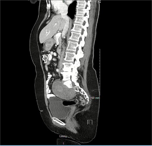
Proximity of schwannoma to rectosigmoid junction on saggital view on repeat scan months later.
The patient re-presented several weeks later for elective resection of her sigmoid colon with concurrent resection of her Schwannoma. The schwannoma was addressed first. Intra-operative neuromonitoring was used to carefully dissect the schwannoma off the retroperitoneum. It was delicately peeled off the iliac vessels and then resected into two pieces (Fig. 3). Attention was then turned to the sigmoid colon. It was dissected in the standard medial-to-lateral approach. The anastomosis was uneventful, and the patient was closed in the standard fashion. She tolerated the procedure well.
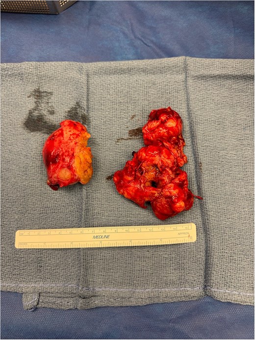
Specimen removed piece-meal.
Post-operatively, the patient had 1 day of right-sided numbness and tingling in her right lower extremity that was self-limiting. Given that this was the side of her schwannoma resection, we opted to observe before ordering any diagnostic studies, such as an EMG. Fortunately, within 24 hours, the patient reported complete resolution of her numbness and tingling. Her hospital visit was slightly prolonged due to rectal bleeding, which was also self-limiting. The patient left the hospital in good spirits.
Discussion
The differential diagnosis for peripheral nerve sheath tumors is broad, encompassing all bPNSTs. Generally, these tumors can be left alone and observed as long as the patient remains asymptomatic. Our patient is unique in the sense that it was not her peripheral nerve sheath tumor that was contributing to her symptoms, but rather her diverticulitis.
The decision to operate and risk damage to surrounding nerve structures, get into uncontrolled bleeding from the iliac vessels, or propagate infection were all taken into consideration before proceeding with surgery. We also discussed that if the tumor were to continue to grow, there could be a slight possibility that a portion of the colon could be compressed secondary to mass effect, ultimately leading to surgery in a more emergent nature. Thus, operative management was chosen over non-operative [7].
Collaboratively, we discussed a staged approach. With colon surgeries, there is always a risk of contaminating the surgical field. This was discussed with neurosurgery and the patient. The patient was adamant that we do this as a combined procedure, as she did not want to wait several months before undergoing another procedure.
To date, there have been no further case reports that describe concurrent schwannoma resection with elective sigmoid resection. It is a feasible operation that requires a multidisciplinary approach from vascular surgery, neurosurgery, and colorectal surgery. At follow-up, the patient was in excellent spirits and had returned to her normal quality of life with no further complications.
Conflict of interest statement
None declared.
Funding
None declared.
