


Abstract
Endometriosis is characterised by endometrial tissue outside the confines of the endometrium in the uterus. Whilst commonly occurring in regions such as the ovaries or fallopian tubes, it is exceedingly uncommon for endometriosis to appear in the umbilical region. This condition may produce a painful and aesthetically unfavourable presentation for sufferers, and can be amendable to surgical excision of the lesion. This case outlines a very rare case of umbilical endometriosis that was managed by an Australian general surgical team.
Introduction
Endometriosis is the presence of endometrial tissue external to the uterine cavity [1]. The peak incidence of the condition is 18–29 years [1]. The condition often causes dysmenorrhea and unpredictable pelvic pain [2]. The goal of treatment is pain management and improved quality of life, which may involve medical or surgical management [2]. The most common site of ectopic tissue is the ovaries, involved in over 60% of cases [3]. Other common areas are the peritoneum, pelvic lymph nodes, cervix, and fallopian tubes [3]. However, the umbilicus is a rare site for endometriosis, accounting for <1% of endometriosis cases [3, 4].
Umbilical endometriosis is either categorized as primary or secondary, with primary lesions occurring in the absence of local surgery, and secondary lesions occurring in scar tissue resulting from surgical skin incisions [4]. Several pathophysiological theories have been suggested to explain primary umbilical endometriosis. These include the transplant theory, the in situ theory, and the induction theory [5]. The transplant theory postulates the migration of endometrial cells via various anatomical routes [4, 5]. The in situ theory described the presence of ectopic cells being due to embryonic cell remnants [5]. The induction theory is a combination of the transplant and in situ theories [5].
We describe a rare and atypical case of primary umbilical endometriosis in a pre-menopausal woman, older than the typical age for presentation. Currently, there are no Australian case reports of umbilical endometriosis.
Case report
A 42-year-old-female was referred to the general surgery outpatient department clinic at a tertiary hospital from the gynaecology clinic. She had originally reported a lesion in her umbilicus to her general practitioner. She complained of 2 years of pain at the site that, interestingly, did not coincide with her menstrual cycles. Approximately 6–12 months after the onset of pain, the lesion at the umbilicus appeared. The patient denied any other symptoms or history of endometriosis. Her only medication was the progestin-only contraceptive pill, and only medical and surgical history was melanoma removal from the chest wall. Her GP had ordered both a biopsy and an ultrasound of the lesion. The biopsy revealed a diagnosis of endometriosis. She was referred to gynaecology who assessed the patient and investigations and expressed concern that excision of the lesion may involve the underlying rectus, hence the referral to general surgery.
On examination, there was a 1–2 cm pink lesion at the umbilicus with multiple nodules. The lesion felt attached to the underlying muscle (Fig. 1). The patient was booked for an excision of umbilical endometriosis.
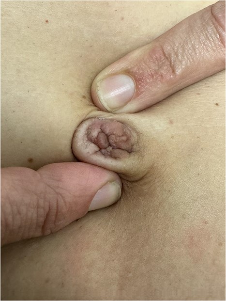
Umbilical endometriosis with suspicion of attachment to underlying rectus.
The procedure was performed under general anaesthetic. An elliptical excision of the nodule was undertaken, with margin down to fascia. The peritoneal cavity was entered, and the fascia was then closed with sutures. An umbilicoplasty was performed and the skin was closed with a vacuum dressing applied.
The procedure was performed as a day case and the patient was discharged without any concerns.
Discussion
Despite endometriosis being a common condition, umbilical endometriosis is exceedingly rare. Even rarer is primary umbilical endometriosis, which occurs in the absence of regional surgical scars or an endometriosis history.
Our patient presented with a non-classical history for primary umbilical endometriosis. She presented at an older age, of 49 years. Furthermore, despite having pain, her pain was not cyclic or coinciding with menstruation. In contrast to other cases, her lesion also did not change in size or bleed during menstrual cycles. It is reported that 90% of primary umbilical endometriosis lesions swell during menstrual cycles [6]. It should be noted that the patient was taking the progestin-only contraceptive pill, which may have had some effect on the clinical presentation.
Imaging can be helpful in ruling out other conditions, and sometimes in assessing the size and depth of the lesion [6]. However, often there is a poor sensitivity and specificity [6]. In our case ultrasound, combined with examination, assisted with risk stratifying the procedure in terms of involvement of the underlying rectus sheath, which was what prompted the referral from gynaecology to general surgery.
Despite a lack of definitive guidelines in the context of the condition’s rarity, primary umbilical endometriosis is often considered an indication for excision by most general surgeons [6]. This conforms to the goal of treatment, being pain management and quality of life, as it removes the source of pain and provides cosmetic relief to the patient. Surgery can also be considered in the context of malignant transformation of the lesion which, although rare, is possible [7].
Due to the rarity of umbilical endometriosis and the poor sensitivity and specificity of imaging, the condition should be considered as a differential diagnosis in females presenting with umbilical lesions. This differential should be considered regardless of the patient’s age, associated endometriosis symptomatology, or previous history of endometriosis. A low threshold for confirmational biopsy should be held and subsequent operative management appears appropriate, with local excision being the mainstay.
Although rare, umbilical endometriosis can occur in the absence of previous endometriosis history or surgery (primary umbilical endometriosis).
The presentation of umbilical endometriosis may be atypical with regards to patient age and symptomatology, with not all cases presenting with cyclical pain and lesion enlargement.
Biopsy is the gold standard of diagnosis with imaging being less helpful, and local excision should be considered to improve symptoms and cosmetics and to reduce the risk of malignant transformation.
Conflict of interest statement
None declared.
Funding
None declared.
