-
PDF
- Split View
-
Views
-
Cite
Cite
Ramez Iseed, Lowry Stanford, Pediatric case of atypical spindle cell/pleomorphic lipomatous tumor within the vastus lateralis muscle, Journal of Surgical Case Reports, Volume 2024, Issue 4, April 2024, rjae219, https://doi.org/10.1093/jscr/rjae219
Close - Share Icon Share
Abstract
This report presents a rare case of an atypical spindle cell/pleomorphic lipomatous tumor in a 13-year-old male patient. The tumor, located within the vastus lateralis muscle, was successfully resected, and the patient is currently under follow-up.
Introduction
Atypical spindle cell/pleomorphic lipomatous tumors (ASPLT) are rare neoplasms that typically present as painless, slow-growing masses. They are often associated with a small risk of local recurrence following marginal excision. These tumors are characterized by ill-defined margins and the presence of variable proportions of mild to moderate atypical spindle cells, adipocytes, lipoblasts, pleomorphic cells, multinucleated giant cells, and a myxoid or collagenous extracellular matrix [1]. ASPLT typically arises from soft subcutaneous tissues of extremities, shoulder, head, neck region, and in the deep soft tissue of extremities. It is unusual in gastrointestinal organs and very unusual in the retroperitoneum tissues. Intra-abdominal ASPLT is mostly seeming to affect mainly men between 45 and 60 years of age.
Case report
A 13-year-old male patient presented with a lump on his left superior thigh, which had been present for several years. The patient reported no pain, tingling, numbness, or tenderness. The patient denied any history of trauma in the region. Past medical history included asthma and ADHD, with no known drug allergies. The patient was up to date with childhood vaccines and had a surgical history of tonsil and adenoid removal.
Physical examination revealed a large mass on the left superior anterior thigh, which was circular, firm, immobile but compressible, and without drainage or erythema.
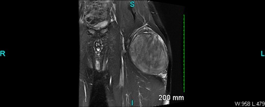
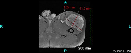
An X-ray of the left femur was negative. An ultrasound revealed abnormal findings, and an MRI with and without IV contrast showed a 10.5 × 7.1 × 11.4 cm heterogeneous enhancing mass within the vastus lateralis muscle without osseous invasion (Figs 1 and 2). There were islands of fat versus hemorrhage within the mass. The overall findings were non-specific but favored a sarcoma.


The patient underwent an excisional biopsy of the left thigh soft tissue mass under general endotracheal anesthetic. A standard limb salvage longitudinal incision over the proximal anterior thigh was performed, and a wedge of tissue was taken for pathology examination. Pathology biopsy report identified the mass and the patient was discharged without complications.
The pathology biopsy report identified the mass as an Atypical spindle cell/pleomorphic lipomatous tumor. This type of tumor does not have metastatic potential but carries a small risk (10%–15%) of local recurrence following marginal excision. The patient was discharged with a prescription for Hydrocodone-Acetaminophen 5–325 mg.
The patient was seen for follow-up 6 months later with no evidence of recurrence and should be seen yearly to prevent local recurrence.
Discussion
This case adds to the limited literature on atypical spindle cell/pleomorphic lipomatous tumors, particularly in pediatric patients. It underscores the importance of thorough investigation and appropriate treatment of soft tissue masses in children. The case presented here is unique due to the patient’s age and the location of the tumor. Most reported cases of ASPLT occur in adults, with a slight male predominance [1]. The tumor in this case was located within the vastus lateralis muscle, which is a less common location.
The case also highlights the importance of imaging in the diagnosis of ASPLT. In this case, an X-ray of the left femur was negative, but an ultrasound revealed abnormal findings, and an MRI showed a heterogenous enhancing mass within the vastus lateralis muscle without osseous invasion. These findings were non-specific but favored a sarcoma [2].
The pathology biopsy report identified the mass as an ASPLT. This type of tumor does not have metastatic potential but carries a small risk (10%–15%) of local recurrence following marginal excision. This emphasizes the need for careful follow-up in these patients.
In conclusion, this case contributes valuable information to the existing literature on ASPLT, particularly in pediatric patients. It highlights the importance of considering ASPLT in the differential diagnosis of soft tissue masses in children and underscores the need for early thorough investigation, appropriate treatment, and careful follow-up in these patients to prevent local recurrence and ensure good patient outcomes.
Conflict of interest statement
None declared.
Funding
None declared.


