


Abstract
Iatrogenic pseudoaneurysms (IPA) are a rare complication of transradial coronary intervention. This brief report aims to present a case report of a radial artery pseudoaneurysm and to review the current management of this entity. Because of the increased performance of transradial coronary intervention, IPA have become more common. Doppler ultrasonography is a fundamental tool to aid the differential diagnosis with other pathologies. There is no consensus on the treatment, which may range from conservative management to surgical repair. Close surveillance after the procedure and early diagnosis are essential to avoid serious complications, such as ischemia of the hand. The management of IPA depends upon morphological characteristics and associated symptoms.
INTRODUCTION
The most common sites for pseudoaneurysm (PA) formation are the femoral and radial arteries, mostly because of iatrogenic causes, such as catheterization for coronary angiography, continuous blood pressure monitoring or arteriovenous shunting for hemodialysis [1]. Less frequently, they may be caused by bone fractures or other traumas or be related to parenteral drug abuse.
When compared with femoral access, transradial coronary intervention is many times preferred, since it provides better comfort for the patient and has a lower complication rate [2]. Despite these benefits, the development of Iatrogenic pseudoaneurysms (IPA) may occur, as well as other complications, such as thrombosis, infection, hemorrhage, ischemia and arteriovenous fistula formation [2]. Transradial approach has a reported incidence of PA of 0.009% [3]. The main difference between a PA and a true aneurysm is the noninvolvement of all three layers of the arterial wall.
The authors present a case of a patient who underwent two coronary angiographies by transradial approach, and then developed a PA.
CASE REPORT
A 62-year-old female with a personal history of atrial fibrillation, hypertrophic cardiomyopathy, heart failure (Classes I–II/IV—New York Heart Association), arterial hypertension, dyslipidemia, Type II diabetes mellitus and smoking underwent two coronary angiography 6 months apart, both through left radial access with a 6Fr sheath and mechanical compression with PreludeSync. Punctures were performed by the Cardiology Department without an ultrasound and without any signs of difficulties in hemostasis. In the last procedure, the patient underwent coronary revascularization with stents and was medicated with dual antiplatelet therapy. The patient developed a pulsatile and expandable mass in the left wrist, 3 weeks after the last coronary angiography (Fig. 1). On physical examination, a painful, expandable and pulsatile mass with a murmur was identified. Complaints of paresthesia or signs of hand ischemia were not present. On duplex ultrasound, the mass had a longitudinal diameter of 2.2 cm, with a narrow neck and turbulent ‘yin-yang’ flow, compatible with a PA of the left radial artery. The patient underwent PA resection with arteriorrhaphy (longitudinal stitches), without any complications, and was discharged 24 h postoperatively (Figs 2 and 3). At 1-month follow-up, the patient was asymptomatic, presenting a palpable radial pulse and did not report any sensory deficits.
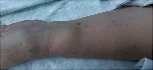
Pulsatile and expandable mass in the left wrist.
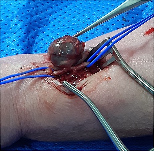
Isolation of the radial artery with the PA.
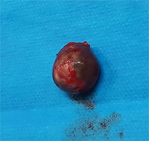
Resected PA.
DISCUSSION
Radial PA are a rare complication, with several predisposing factors such as multiple puncture attempts, systemic anticoagulation, inadequate hemostasis, vascular site infection, and the use of larger sheaths which may lead to the development of this type of PA [4]. In order to reduce the risk of complications from transradial approach for coronary procedures, some measures such as a single puncture, effective compression and early surveillance, are recommended [4].
On physical examination, a radial artery PA may present as a pulsatile mass, associated with local pain and hand edema. Neuropathy or ischemia of the hand might be present, depending on dimension of the mass [4]. Doppler ultrasonography is a fundamental tool to aid the differential diagnosis with other pathologies, such hematoma, abscess or a true aneurysm. On Doppler ultrasound, the ‘yin-yang’ sign indicates bidirectional flow because of the swirling of blood within the PA.
Regarding treatment, because of a few number of cases published, no uniform consensus has been established [5]. A conservative approach can be performed in asymptomatic patients in the presence of a small PA (i.e. <10 mm in diameter), through ultrasound guided compression or minimally invasive thrombin injection have also been employed [1]. However, compression with a probe is user-dependent and sometimes inefficient in patients usually on multiple antithrombotics [6]. Thrombin injection is a valid option for the treatment of narrow neck PA in other locations; however, radial PA carries the risk of embolizing thrombin to the digital arteries leading to ischemia of the fingers and necrosis. Beyond this risk it does not eliminate the mass effect and extrinsic compression on nervous structures, nor skin tension which may ultimately lead to deleterious effects.
Regarding endovascular techniques, embolization or endovascular exclusion of the PA with covered stent (by brachial approach) followed by decompression with a syringe is an option that has already been successfully described, but needs further studies [7].
Surgical treatment is suggested for large PA (>10 mm in diameter), fast-growing PA, infected PA or in cases of significant mass effect, such as ischemia of the hand, neuropathy and soft tissue necrosis. Surgical repair is also indicated when conservative treatment fails. There are several validated surgical techniques, such as radial artery ligation with PA resection if cubital artery perfusion and palmar arch are not impaired, primary suture repair, end-to-end reconstruction or graft interposition.
In the case described, the resection and primary repair of the radial artery defect was chosen taking into account the size of the PA and the mass effect produced along with the relatively restricted arterial wall lesion.
To conclude, because of increased transradial approach in coronary procedures, radial IPA are becoming more frequent. Close surveillance and early diagnosis of PA’s are essential to avoid serious complications. Treatment may be conservative or surgical depending on the morphological characteristics and associated symptoms of the PA.
ACKNOWLEDGEMENTS
Angiology and Vascular Surgery Department, Hospital do Divino Espírito Santo (HDES), Ponta Delgada, São Miguel Island, Azores, Portugal.
CONFLICT OF INTEREST STATEMENT
None declared.
FUNDING
This case report received no specific grant from any funding agency in the public, commercial or not-for-profit sectors.
DECLARATION OF PATIENT CONSENT
The authors certify that they have obtained all appropriate patient consent forms. In the form, the patient has given his consent for his images and other clinical information to be reported in the journal. The patient understand that his name and initials will not be published, and due efforts will be made to conceal his identity.
