-
PDF
- Split View
-
Views
-
Cite
Cite
Kyle J Klahs, Ethan Heh, Mohammad Yousaf, Joshua Tadlock, Ahmed M Thabet, Operative challenges of intramedullary nailing for subtrochanteric blastic pathological femur fracture: a case report, Journal of Surgical Case Reports, Volume 2023, Issue 1, January 2023, rjac630, https://doi.org/10.1093/jscr/rjac630
Close - Share Icon Share
Abstract
Prostate adenocarcinoma metastasizes to bone and forms fragile blastic lesions, which can present as dense obstacles intraoperatively. There are limited reports on the challenges surgeons face when operating through these lesions. A 60-year-old male with a pathologic subtrochanteric femur fracture in the presence of blastic lesions was successfully treated with intramedullary (IM) fixation. Pathologic fractures from blastic bone lesions are expected to increase in prevalence as survivability improves for metastatic prostate cancer. Orthopedic surgeons, when performing IM fixation for these fractures, should be prepared to utilize accessory equipment and should adopt creative techniques for reduction and fixation.
INTRODUCTION
Prostate cancer most commonly metastasizes to and forms blastic bone lesions [1]. The process is multifactorial but involves osteoclastogenesis downregulation from prostate-specific antigen promoting osteoprotegerin [2, 3]. Risk of fracture is increased 1.9-fold and typically occurs in proximal long bones [1, 4]. Intramedullary (IM) is the preferred fixation for subtrochanteric femur fractures; however, osteoblastic lesion may complicate the IM trajectory [5–7]. There are few reports of intraoperative complications when using IM, and only 1% was documented in a review by Ormsby et al. [8]. This study will describe a case of intraoperative challenges due to the osteoblastic lesions encountered during IM fixation of a subtrochanteric femur fracture.
CASE REPORT
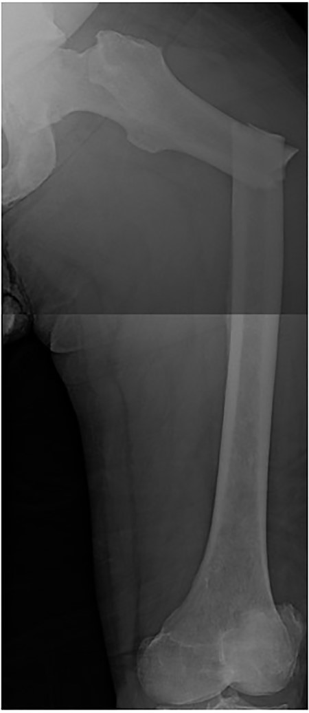
A 60-year-old male, who was diagnosed with Stage 4 prostate adenocarcinoma with known rib metastases and treated with chemotherapy and prostatectomy, sustained a pathologic left transverse subtrochanteric femur fracture after a ground-level fall while ascending stairs at his home. He denied any antecedent left hip or thigh pain. Radiographs revealed blastic metastatic lesions to his left femur (Fig. 1).
Preoperative planning
We assembled a multidisciplinary team of medical, surgical and ancillary professionals. Preoperative labs and radiographs to include contralateral full-length femur XRs were obtained to investigate metastatic lesions to the right femur. The right proximal femur demonstrated peritrochanteric blastic lesions <one-third of the diaphyseal diameter (Mirel’s score = 5) [9]. Our patient was estimated to have over a 12-month survival, making him an ideal surgical candidate.
Intraoperative
The patient was positioned supine on a fracture table. A guide wire was inserted through a 4-cm surgical incision proximal to the greater trochanter (GT) and was passed through a cannulated awl in a position slightly medial to the tip of the GT on the AP and center on the lateral (Fig. 2B). A (15-mm) entry reamer widened the opening (Fig. 2C). A rigid cannulated reduction rod and forceful malleting allowed the ball-tipped guidewire to cross the close reduced fracture, but too lateral and posterior distally (Fig. 3A and B). The cannulated flexible reamers encountered impassible blastic lesions within the proximal femur (Figs 3C and 5B).
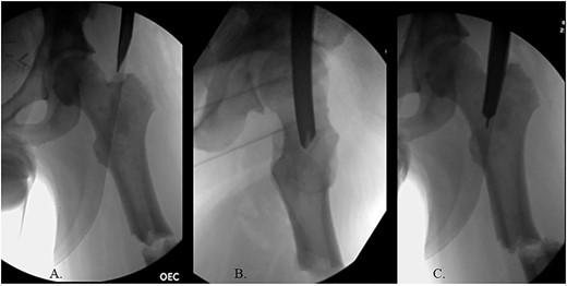
(A) Cannulated awl positioned medial to tip of GT on the AP XR; (B) cannulated awl positioned center of GT on the lateral XR; (C) entry reamer over guide pin on the AP XR.
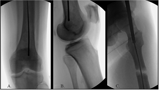
(A) Ball-tipped guidewire slightly lateral position at the knee on the AP XR; (B) ball-tipped guidewire too posterior at the knee on the lateral XR; (C) flexible reamer within the proximal femur abutting blastic lesions.
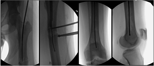
(A) Canulated cutter tool creating a new path in the proximal femur; (B) rigid canulated reduction rod directing the guidewire across a reduced fracture held with a proximal bone hook and distal ball spike pusher; (C) center positioned ball-tipped guidewire at the knee on the AP XR; (D) center positioned ball-tipped guidewire at the knee on the lateral XR.
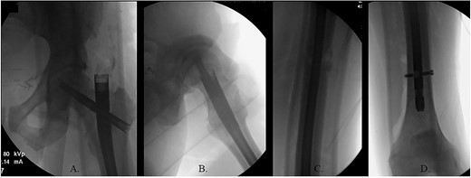
(A) AP radiograph of left hip with implant; (B) lateral radiograph of left hip with implant; (C) reduced fracture site with implant; (D) distal femur with implant.
The ball-tipped guidewire was extracted, and a sharp cannulated cutter created a novel path in the proximal segment (Fig. 4A). A rigid nonunion reamer (DePuy Synthes©, Raynham, MA) was advanced to widen the canal. Closed reduction was lost and so a lateral incision was made at the level of the fracture and a percutaneous reduction was achieved through use of a proximal bone hook and distal ball spike pusher (Fig. 4B).
The hip was extended, and a ball-tipped guidewire was successfully passed across the fracture site to a center–center positionwithin the canal at the level of the knee (Fig. 4C and D). Sequential 0.5-mm reaming from 9 to 13 mm prepared for an 11 × 400 mm, 125° Gamma3® intertrochanteric rod (Stryker© Kalamazoo, MI), with a 95-mm cephalomedullary screw and ×2 distal lateral to medial interlocking 5.0-mm screws (Fig. 5A–D). Through the course of the procedure, 400 cc of blood loss necessitated two units of packed red blood cells.
Post-operative
The patient immediately was weight-bearing as tolerated to the operative extremity and worked with physical therapy (PT) to include 80 ft on post-operative day (POD) #2 with use of a front-wheeled walker. He continued to progress with PT and was discharged home POD #8 with home health/PT. At 12 months, the patient denied pain, and XRs demonstrated robust callus formation and bridging healing at the fracture site (Fig. 6).
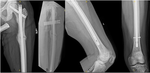
(A) 12-month post-operative AP radiograph of left hip; (B) 12-month post-operative lateral radiograph of left hip; (C) 12-month post-operative lateral femur radiograph; (D) 12-month post-operative AP distal femur radiograph.
DISCUSSION
With increasing prevalence due to extended survivability, orthopedic surgeons will be treating more pathologic fractures secondary to prostate adenocarcinoma metastasis [10–13]. These blastic lesions alter the internal bone morphology by increased cortically dense obstacles, providing challenges for the surgeon.
Treatment options for pathologic subtrochanteric femur fractures include open reduction internal fixation (ORIF), endoprosthetic reconstruction and IM, with a preference for IM due to the minimal surgical approach as compared to ORIF and the patient’s ability to weight bear immediately after surgery [5, 6, 14, 15]. Maximizing IM implant length can also protect the entire bone from expanding or noncontiguous lesions [16, 17]. Intralesional reaming for IM fixation with post-operative adjuvant multifractionated radiation therapy is an acceptable palliative option in known metastatic lesions [18–21]. A systematic review by Janssen et al. reviewed 40 studies on proximal femur metastatic fixation and determined that the reoperation rate were significantly less for endoprosthesis and IM as compared to ORIF, whereas deep infection was greatest in the endoprosthesis group [22].
The IM fixation of subtrochanteric fractures can present as more difficult than proximal intertrochanteric femur fracture or distal femur shaft fractures due to the muscular attachments on the proximal segment and their resultant deforming forces [23]. Adding blastic lesions obstacles to the implant trajectory necessitates utilization of other tools such as the hand-cutting femur nonunion reamer to bore out a path for the flexible reamers to follow. Blastic lesions, although dense are brittle, and therefore, care must be taken to avoid the propagation of the fracture or additional iatrogenic fractures, making incremental reaming paramount [24].
As the incidence of metastatic blastic pathologic fractures increase, the orthopedic surgeon should be prepared to treat these difficult fractures. Utilizing equipment from nonunion trays as well as percutaneous reduction techniques can provide an advantage in successfully and safely preparing the femur for an IM device.
STATEMENT OF INFORMED CONSENT
The patients were informed that data concerning the case would be submitted for publication. The patients agreed and consented to release of this data for publication.
CONFLICT OF INTEREST STATEMENT
None declared.
FUNDING
None.


