


Abstract
Oesophageal squamous papillomas (OSPs) are rare epithelial lesions, recognized to be benign but with reported malignant potential. We report a case of a 40-year-old female with chronic vomiting, subsequently found on oesophago-gastro-duodenoscopy to have two mid-oesophageal sessile polyps, the largest of which measured 10 mm. These were endoscopically resected with histopathology confirming an OSP without evidence of dysplasia or malignancy. The case under consideration reports the current literature on OSPs regarding their aetiology, malignant potential and optimal management.
INTRODUCTION
Oesophageal squamous papillomas (OSPs) are rare epithelial lesions, recognized to be benign but with malignant potential. We report a case of an adult female found to have two mid-oesophageal sessile polyps on oesophago-gastro-duodenoscopy (OGD). These were endoscopically resected with histopathology confirming OSP without evidence of dysplasia or malignancy. The case considers the current literature on OSPs regarding their aetiology, malignant potential and consequent management.
CASE REPORT
A 40-year-old woman was diagnosed with a protracted history of multiple episodes of dry retching and vomiting each day over 4–5 years. Her co-morbidities included hypertension requiring a β-blocker, an angiotensin receptor blocker, and she was a current smoker. Clinical examination was unremarkable. Computed tomography imaging of the abdomen was normal.
The patient underwent an OGD with findings of two sessile polyps in the mid-oesophagus, the largest measuring 10 mm (Fig. 1). Both lesions were endoscopically resected and subsequent histopathology demonstrated oesophageal squamous cell papillomas (OSPs) without evidence of dysplasia or malignancy.
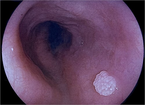
Endoscopic view of a 10-mm polypoid sessile lesion in the mid-oesophagus.
DISCUSSION
Oesophageal squamous cell papillomas are rare and benign epithelial lesions, occurring in 0.01–0.45% of persons undergoing OGD [1–4]. Although OSPs have been reported in almost every age group, the majority are diagnosed in the fourth to sixth decades of life and with a predilection for females [1–5]. Most are asymptomatic and found incidentally during OGD for other indications; however, associated symptoms of dyspepsia, pyrosis, epigastric discomfort and dysphagia may be present [1–4].
OSP are commonly found in the middle and distal thirds of the oesophagus as solitary and small lesions ranging between 2 and 6 mm in size, although they have been found in all parts of the oesophagus [1–5]. Lesions greater than 10 mm, clusters of lesions and diffuse involvement of the oesophagus have been reported in the literature but are rare [6–8]. Endoscopic findings are of a fleshy white-pink coloured lesion with wart-like exophytic projections [2]. Although these macroscopic features are not pathognomonic for OSP, the triad of an exophytic growth, wart-like projections and surface vessels visualized on narrow band imaging as crossing the lesion were found to have a positive predictive value of 88% in one study [2]. Differential diagnoses include verrucous squamous cell carcinoma, malignant melanoma, leiomyoma, papillary leucoplakia and inflammatory fibroid polyp. On histopathology, OSPs are characterized by a fibrovascular core of connective tissue and small blood vessels branching out from the lamina propria, and lined by acanthotic stratified squamous epithelium, without dysplastic or neoplastic changes [2, 8].
The exact aetiology and pathogenesis of OSP remains uncertain. Chronic mucosal irritation with hyper-regenerative responses, together with human papillomavirus (HPV), given its role in genital and oropharyngeal warts and cancers, have been implicated [2]. The role of gastro-oesophageal reflux is supported by observations of OSP arising in the distal oesophagus where chronic irritation with refluxing gastric acid occurs [1]. Other chemical and mechanical factors such as caustic injury, nitrosamine exposure, smoking, alcohol, prolonged nasogastric intubation, and also previous oesophageal dilatation, have been associated with OSP [7]. HPV has been detected in OSP in several studies, with a prevalence of 10–80% reported in the literature [4, 5, 8, 9]. However, the HPV serotypes tested in most studies were either not stated or limited to HPV 6, 11 and 16 such that the association of HPV and OSP may be under-estimated due to the large number of existing HPV serotypes not routinely tested [3, 5, 9].
Malignancy arising from OSP is rare, with only a small number of cases complicated by squamous cell carcinoma (SCC) having been reported in the literature [3, 4, 8, 10]. Features suspicious for malignant potential include the presence of multiple lesions, papillomatosis or large lesions, although thresholds are yet to be determined, given the rarity of cases [3, 8, 10]. HPV infection may be responsible for malignant transformation due to the known role of high-risk subtypes in cervical, anogenital and oropharyngeal SCCs [5]. This, however, remains to be established as HPV was not detected in the few reported cases of OSP complicated by malignancy, but again the serotypes tested were either limited to low-risk HPV 6 and 11 in one study, HPV 6, 11, 16, 18, 31, 33 and 35 in another or not stated [3, 8, 10].
Long-term outcomes and prognosis are largely unknown due to the rarity of OSP and the paucity of information in the literature [4]. The current consensus is that OSP should be resected completely given the small but real risk of malignant potential, and the improvement of symptoms in symptomatic cases [6, 8, 10]. Techniques include forceps biopsy resection, snare polypectomy, cautery or radiofrequency ablation for small, solitary lesions and endoscopic mucosal resection, endoscopic spray cryotherapy or indeed oesophagectomy for more extensive OSPs [1, 3, 7].
In conclusion, OSP is rare and most often found incidentally on OGD performed for other indications. They can be easily missed due to morphology and location, hence vigilance is key. Future research should aim to elucidate the aetiology and natural history of OSPs, and to include the predictors of malignant potential, so as to optimize management and surveillance strategies.
CONFLICT OF INTEREST STATEMENT
None declared.
