-
PDF
- Split View
-
Views
-
Cite
Cite
Jessica Saifee, Mackenzie Shindorf, Omar Samara, Steven Bourland, Stig Somme, Incarcerated umbilical hernia in a 22-month-old child, Journal of Surgical Case Reports, Volume 2022, Issue 3, March 2022, rjac052, https://doi.org/10.1093/jscr/rjac052
Close - Share Icon Share
Abstract
Incarceration of an umbilical hernia (UH) rarely occurs in the pediatric population. They usually resolve spontaneously or are treated after the child turns 4–5 years old [1, 2]. Risk factors for incarceration have been identified, but little is understood about how incarceration of an UH occurs.
INTRODUCTION
An umbilical hernia (UH) is a protrusion of the abdominal viscera through the umbilical ring [3]. UHs typically occur in children due to the failure of the umbilical ring to close, but usually resolve spontaneously by the age of 4 [4]. Incarceration of a pediatric umbilical hernia is rare, occurring at a rate of ~1 in every 1500 [2]. Risk factors for pediatric umbilical hernia occurrence include being premature, a low-birth weight, hypothyroidism and African American [5–7]. This report describes the case of a young 22-month-old African American female with an incarcerated umbilical hernia secondary to foreign bodies.
CASE REPORT
A 22-month-old girl was admitted to the hospital with abdominal pain, nausea, vomiting and fatigue for 2 days. She was initially seen at an outside emergency room where imaging studies of the abdomen were obtained. According to the mother of the child, she was playing outdoors and had consumed dirt, grass and small rocks.
Physical exam demonstrated a child who was hemodynamically stable and uncomfortable appearing with a soft, but distended abdomen, that was tender in the area of a prominent umbilical hernia, that was not reducible.
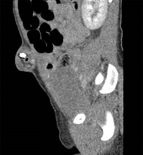
Her laboratory results demonstrated a leukocytosis with a left shift 11.27. Abdominal x-ray and CT imaging studies from the outside ED were reviewed and showed bowel obstruction with radiopaque objects near the neck of the hernia (Fig. 1). Attempts at manual reduction of the umbilical hernia were unsuccessful and the decision was made to proceed to the operating room for an umbilical exploration.

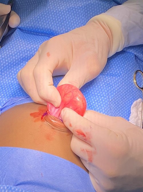
Intraoperatively, the umbilical hernia sac was opened to evaluate and reduce the bowel (Fig. 2). Within the incarcerated loop of small bowel, the rocks and dirt were palpated. The incarcerated small bowel was successfully reduced. The entirety of the small bowel was subsequently evaluated for additional foreign bodies, and more specifically rocks and dirt. All areas of the small bowel were found to be viable and multiple small stones were palpated but none were obstructing the lumen. A 3 cm fascial defect was identified and closed primarily with a 2-0 vicryl.

DISCUSSION
UH is a common occurrence among the pediatric population. The rate of incarceration in the general population is reported to be 1 in 1500 [8]. Although, this number could be smaller since the exact incidence of asymptomatic umbilical hernia is unknown.
We believe that the incarceration of the small bowel within the hernia sac was due to the ingested stones not being able to pass back through the fascial defect and subsequently caused a small bowel obstruction [6, 9].
This case is also the first case in reported literature where the cause of incarceration included rocks and dirt; other reported causes included Meckel’s diverticulum, open safety pin digestion, and gangrenous retrocolic appendix [10]. This reported case was rare given the size of the umbilical hernia and cause.
One of the most common causes of incarceration is omental incarceration, especially in smaller umbilical defects, this is less urgent to address surgically but can cause significant pain [11].
Although incarceration of an umbilical hernia is rare, it is important to monitor them for potential complications, which include abdominal pain, nausea, and vomiting. Incarcerated UH require prompt surgical intervention to prevent ischemic damage.
INFORMED CONSENT
No PHI was included in this report.
CONFLICT OF INTEREST STATEMENT
None declared.
FUNDING
No funding sources to report.


