


Abstract
Acute Type-A Aortic Dissection is a rare but life-threatening surgical emergency in children and adolescents. Large series report up to 3.5% of cases under the age of 21 with many of these cases with known predisposing risk factors. Risk factors include congenital cardiovascular disorders, connective tissue disorders, hypertension, inflammatory aortic weakening and iatrogenic, pregnancy or trauma. Cases with no obvious predisposing risk factors are exceedingly rare with very few cases described in a limited number of studies.
INTRODUCTION
Stanford Type-A Aortic Dissection in children and young adults is rare with an incidence of 0.67–3.5% in patients under the age of 21 [1]. It is associated with high mortality, and several risk factors have been implicated including congenital cardiovascular disorders, connective tissue disorders, hypertension, inflammatory aortic weakening and iatrogenic, pregnancy or trauma. Aortic dissection in pediatric patients with hypertension or no identifiable risk factors is exquisitely rare and has only been described in a limited number of studies [1, 2]. Studies on aortic dissection and the Australia Indigenous Population are scarce. The rarity of this disease within this patient demographic translates into diagnostic difficulties and repercussions on timely treatment with mortality rising 1–3% per hour without surgical intervention [2–5]. This case report describes the rare case of a 15-year-old Indigenous male presenting with Acute Type-A Aortic Dissection and no pre-existing risk factors.
CASE REPORT
A 15-year-old Australia Indigenous male presented to the pediatric emergency department with severe sudden onset central chest pain, difficulty breathing and diaphoresis. This patient suffered from autism spectrum disorder and fell into greater than 95th percentile for weight. His parents had noticed increased levels of agitation two hours prior to presentation to the emergency department with non-verbal communication pointing towards the patient’s own chest, abdomen and neck. The pain was unprovoked, and he had no known pre-disposing risk factors.
The patient was hemodynamically stable during his admission; however, worsening pain profile led to a serial electrocardiogram showing sinus rhythm with ST segment changes consistent with ischemia. There was an initial troponin rise of 245 ng/L progressing to a peak of 922 ng/L. The remaining blood panel was unremarkable. Chest X-ray showed no widened mediastinum and clear lung fields. A bedside transthoracic echocardiogram demonstrated a dissection flap with severe aortic regurgitation in the ascending aorta. Computed tomography aortogram was consistent with a Stanford Type A aortic dissection extending from the aortic root to the bifurcation of the common iliac arteries (Figs 1 and 2). There was involvement of the right brachiocephalic artery and right common iliac.
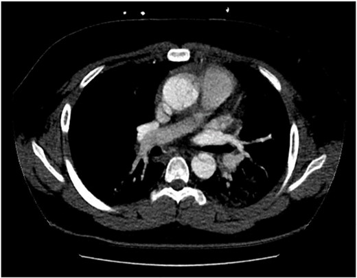
Computed tomography aortogram Axial Image of Stanford Type-A Aortic Dissection.
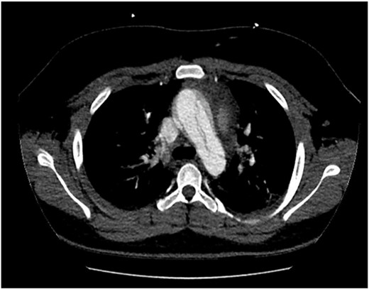
Computed tomography aortogram Axial Image of Stanford Type-A Aortic Dissection extending into arch and descending thoracic aorta.
The patient underwent a Bentall’s Procedure utilizing a 25 mm St Jude Mechanical Valve. Time from presentation to theatre was 9 h. Total length of hospital stay was 10 days of which 5 days were spent in the pediatric intensive care unit. Histological features of the aortic valve and aorta were consistent with cystic medial degeneration. Screening tests were unremarkable for both the patient and close kin. Computed tomography imaging of close kin revealed normal aortic root and ascending aorta dimensions.
DISCUSSION
Type-A Aortic Dissection in children and adolescents is unfamiliar territory amongst even experienced clinicians with limited literature available to improve awareness and understanding. Subsequent delay in diagnosis and definitive treatment translates to high hospital morbidity and mortality.
Shamszad et al. and Fikar et al. are the largest epidemiological studies available in aortic dissection in patients under the age of 21 with the remaining data generally limited to small patient series or case reports [1, 2, 5]. Congenital cardiovascular disorders and trauma were the most common predisposing risk factors with connective tissue disorders and hypertension less common. Although majority of aortic dissection in children and young adults demonstrate predisposing risk factors, 22% of patients had no apparent risk factors [1–3]. There is predominance to male obese patients [3]. Cases of Type-A Aortic Dissection in patients 15 years of age or younger without known risk factors are extremely rare as evident in this case.
Rarity, atypical presentation and normal initial parameters may affect triage categorization and delay in diagnosis. Increasing intensity of pain, agitation and clinical deterioration prompted a serial electrocardiogram and troponin evaluation and subsequent echocardiography demonstrating an aortic dissection flap and severe aortic regurgitation. Contrast-enhanced computed tomography is usually the preferred imaging modality in the diagnosis of aortic dissection; however, this can be difficult to do in younger patients and especially those with behavioral disorders [3–5]. Cultural and communication barriers specific to this case contributed to the diagnostic challenge and exemplify the importance of thorough and repeated clinical examination in this age group.
Type-A Aortic Dissection without pre-disposing risk factors remains a rare but known presentation in children and adolescents. Patients in this demographic who present with symptoms concerning for acute aortic syndrome, especially those who are male and obese, should prompt investigation with transthoracic echocardiogram and/ or computed tomography imaging irrespective of risk factor status. Delayed or misdiagnosis has deleterious effects on hospital morbidity and mortality; therefore, prompt surgical intervention is paramount. Screening tests for the affected patient and family members is recommended including computed tomography evaluation of aortic root and ascending aorta.
DATA AVAILABILITY
The data that support the findings of this study are available from the corresponding author, AR, upon reasonable request.
CONFLICT OF INTEREST STATEMENT
None declared.
FUNDING
None.
