


Abstract
Acute appendicitis is one of the most common causes of acute abdominal pain globally. The pathophysiology of acute appendicitis is due to occlusion of the appendiceal lumen commonly from a faecolith. Obstruction of the appendiceal lumen by ingested foreign bodies is possible albeit rare. Here, we present an extremely rare case of acute appendicitis caused by impaction of the patients tooth within the lumen of the appendix. There have been only seven reported cases of impacted teeth causing appendicitis in the literature. There are no evidence-based guidelines for the management of appendicitis caused by tooth impaction. The authors suggest operative management with appendicectomy should be considered in the first instance.
INTRODUCTION
Acute appendicitis is one of the most common causes of the acute abdomen in adults. The lifetime risk of appendicitis is 8.6% for males and 6.7% for females [1]. Generally, the pathophysiology of appendicitis is thought to be due to luminal obstruction, most commonly from an impacted faecolith. The resulting production of mucus by mucosal goblet cells and gas by luminal bacteria results in distension of the distal appendix causing pain and inflammation. Impaired venous return can progress to ischaemia and perforation [2]. We present an extremely rare case of septic shock from acute appendicitis caused by an impacted molar tooth.
CASE PRESENTATION
A 52-year-old gentleman who presented to the Emergency Department in septic shock with right sided abdominal pain. He suffered a witnessed cardiac arrest prompting immediate cardiopulmonary resuscitation (CPR) with return of spontaneous circulation (ROSC) after 10 minutes. Physiologic resuscitation was commenced, and he was started on broad spectrum intravenous antibiotics with cefuroxime and metronidazole, as per local guidelines. Following intubation and ventilation, he underwent a computer tomography (CT) scan of the abdomen which revealed moderate volume of free fluid with locules of gas within the pelvis suggesting a hollow viscus perforation. A calcified structure was identified within the appendix raising the possibility of a foreign body causing perforated appendicitis. On reconstructed slices, the appearance was that of a tooth (Figs 1 and 2).
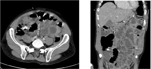
Axial and coronal CT slices showing radiopaque object in the right lower abdominal quadrant.
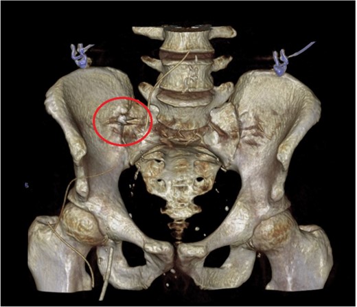
CT reconstruction showing a tooth near the pelvic brim.
The patient proceeded immediately for a laparotomy. Intra-operative findings were of perforated appendicitis with four-quadrant purulent peritonitis. The caecum was healthy and an appendicectomy was performed. On examination of the appendix specimen, there was a perforation in the mid-point and a firm foreign body causing luminal obstruction at the base. On opening the specimen, the foreign body was identified as a decaying tooth containing a metallic filling (Fig. 3).
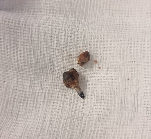
Specimen photograph of the retrieved tooth containing a metallic filling.
Following washout with copious irrigation, the patient’s abdomen was closed and transferred to the intensive care unit (ICU). Unfortunately, over the next 36 hours, he passed away secondary to significant multi-organ failure related to sepsis.
DISCUSSION
The pathophysiology of appendicitis is thought to be due to obstruction of the appendiceal lumen and the cause of luminal obstruction of the appendix is most commonly a faecolith. The prevalence of faecolith at appendicectomy has been reported widely to be between 1.5 and 65.1% [3–5]. Obstruction of the appendix lumen due to an exogenous foreign body is extremely rare in the adult population. In particular, there have been only seven cases of ingested teeth and dental prostheses causing appendicitis in the literature [6–11].
Antibiotic therapy as an alternative to appendicectomy has long been an area of interest in literature. Some recent high-powered studies have added weight to this treatment choice. However, these studies show that participants with faecolith had significantly higher rates of appendicectomy compared to those without faecolith and had higher rates of complication, 20.2 vs 3.6 per 100 [12]. Similarly, appendicitis caused by a foreign body is not likely to resolve with conservative treatment with antibiotics alone due to the continued mechanical obstruction of the appendix lumen. In particular, foreign bodies causing appendicitis often cause perforation [13] and complicated appendicitis is not amenable to conservative treatment with antibiotics alone.
This case highlights the risk of significant morbidity and mortality due to appendicitis due to foreign bodies and dental prostheses. There are no evidence-based recommendations tha determine the best approach to both diagnosis or treatment appendicitis caused by tooth impaction however the authors suggest it is reasonable to treat them in the same way as faecoliths or other foreign bodies with appendicectomy in the first instance.
CONFLICT OF INTEREST STATEMENT
None declared.
FUNDING
None.
References
Simkovic D, Hladík P, Lochman P.
