


Abstract
An adolescent presented with a 5 hour of history of unilateral testicular pain and examination findings in keeping with a unilateral testicular torsion. On scrotal exploration, there was evidence of bilateral testicular torsion and bilateral orchidopexy was subsequently performed. Intravaginal bilateral testicular torsion is scarcely reported in adolescents and adults. Both unilateral and bilateral testicular torsions in adolescents are commonly associated with anatomical anomalies, which were not evident in this case. Despite warming the symptomatic testis for 40 minutes, viability was indeterminate. Guidelines remain equivocal about adequate reperfusion period before considering orchiectomy. Interval sonographic follow-up is recommended to assess testicular atrophy; however, considerable variation exists in practice. This is an atypical case of bilateral synchronous testicular torsion demonstrable by an absence of classical risk-factors, alongside unilateral clinical signs and symptoms. Inconsistency in guidelines for attempted reperfusion could result in orchiectomy where testicular salvage is possible.
CASE REPORT
A 15-year-old male presented with a 5-hour history of sudden onset right hemi-scrotal pain. The pain started during sleep with no precipitating factors or history of trauma. The patient had no past medical history and denied any previous sexual history. The patient was heavy for his age, weighing 84 kg. On examination, the right testicle was high-riding, hard and tender with an absent cremasteric reflex. Examination of the left testicle was unremarkable, with normal size, lie and cremasteric reflex.
The patient was taken to theatre for urgent scrotal exploration 8 hours following onset of initial symptoms. Exploration of the right hemi-scrotum revealed a 180° clockwise intravaginal twist with the right testis appearing blue and dusky with reactive hydrocele. The testicle was delivered and detorted with no immediate change to colouration, prompting attempted reperfusion. The left hemi-scrotum was explored which revealed a non-torted cord, however, similar dusky blue appearance of the left testis which was then warmed in the same fashion. Following 20 minutes, the left testicle appeared viable and orchidopexy was performed. The symptomatic right testis, after a total of 40 minutes, still appeared dusky but improved. A right sided orchidopexy was thus performed.
The patient recovered well post-operatively and was discharged the following day. Follow-up after 3 months with an ultrasound scan revealed symmetrical well-preserved vascularity within both testes (Fig. 1).
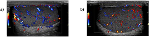
Sonographic appearances of (a) right testis and (b) left testis with colour doppler demonstrating adequate perfusion.
DISCUSSION
Testicular torsion is a urological emergency, occurring at a rate of 1 in 4000. It is characterized by twisting of the spermatic cord and vascular compromise to the testicle [1]. A bimodal distribution has been recognized in two age groups: adolescents and neonates.
Bilateral torsion is a rarer entity with fewer cases described in the literature [2, 3]. The vast majority are cases of extravaginal torsion in neonates. Adolescents comparatively demonstrate intravaginal torsion commonly associated with the ‘bell clapper deformity’; an abnormally high attaching tunica vaginalis on the spermatic cord, which is bilateral in 66–100% of cases [4]. Other risk factors for torsion have been identified, including cryptorchidism, trauma, cold weather, exercise and sexual activity. Preservation of fertility necessitates urgent scrotal exploration, the gold standard investigation and treatment of torsion, within 6 hours from onset [5]. Following detorsion, the salvaged and contralateral testes undergo orchiopexy to prevent retorsion; however, recurrence after orchiopexy occurs in 4.5% [7].
This case highlights several key points for discussion. Firstly, while duration of torsion is a well-known predictor of testicular viability [5], limited information is available regarding the optimal time to wait for reperfusion of the testis. The literature recognizes factors favouring potential viability including arterial bleeding on incision of the tunica albuginea, prompt coloration following detorsion, low degree of twisting and short duration of symptoms (<6 hours) [1, 8]. The right testicle of our patient was not detorted until 8 hours from symptom onset, nor did it demonstrate prompt reperfusion. However, following 40 minutes of warming, signs of reperfusion were evident, and after fixation, vascularity confirmed on ultrasound after 3 months (Fig. 1). The lack of clear guidance regarding the time to wait for reperfusion could lead to potentially viable testis needlessly being excised. In bilateral torsion, the consequences of excising potentially salvageable testes would lead to irreversible infertility and androgen deficiency. Conversely, salvaging even one testis preserves endocrine function [8] and the potential for future fertility; albeit with abnormal sperm morphology, low motility and reduction in sperm count [9].
The need for contralateral hemi-scrotal exploration is re-emphasized. Our patient presented with unilateral right sided torsion with a low index of suspicion for left testicular pathology; however, on scrotal exploration, the left testicle also demonstrated ischaemic changes without signs of twisting, perhaps alluding to intermittent testicular torsion requiring orchidopexy. Remarkably, this patient had no anatomical variants predisposing to torsion; hence, the absence of these features should not preclude bilateral exploration as unilateral torsion alone is a risk factor for contralateral torsion [10]. The lack of clinical signs in the left hemi-scrotum demonstrates the diagnostic uncertainty associated with the acute scrotum, perhaps the overwhelming pain in the right testis masked symptoms in the left testis. Benge et al. (1992) [11] reported a case of an 11-year-old boy with presumptive right testicular torsion and an unremarkable left hemi-scrotum, who later demonstrated a 720° right and 360° left testicular torsion. Bilateral scrotal exploration is therefore a necessity.
The main limitation of this case study is the short duration of follow up (3 months). Evidence suggests that many testes deemed ‘viable’ at the time of scrotal exploration may undergo atrophy on long-term follow-up. Lian et al. (2016) [12] show in a study of 37 patients with ‘salvaged testes’, 20 (54%) developed testicular atrophy at a mean duration of 6 months (range 2–14 months). Thus, while our patient had vascularity confirmed on follow up, it remains to be seen whether both testicles may later undergo atrophy and the implications this may have on future fertility. Current guidelines support a 6-month follow-up [13]; however, variation exists between trusts with some advocating 6–8 weeks [14]. Aside from detection of testicular atrophy, follow-up at 6 months is of value in the provision of psychological support, gauging sub-fertility and in considering adolescents for hormone replacement therapy to aid pubertal development [9].
CONCLUSION
We report a unique case of bilateral synchronous torsion with an absence of classical risk factors, atypical presentation and delayed reperfusion following detorsion. This case highlights the diagnostic uncertainty facing urologists in testicular torsion and the importance of bilateral scrotal exploration.
Current British Association of Paediatric Urologist guidelines do not explore the time to wait for reperfusion following detorsion or intra-operative factors favouring salvage in testicular torsion [14]. This may lead to heterogeneity in the rates of orchiopexy versus orchiectomy between centres, resulting in the excision of potentially viable testes. Follow-up at 6 months is re-emphasized to adequately manage the complications of testicular torsion.
