-
PDF
- Split View
-
Views
-
Cite
Cite
Andreas Skarpas, Petros Siaperas, Athanasios Zoikas, Emmanouela Griva, Ioannis Kyriazis, Georgios Velimezis, Ioannis Karanikas, Meckel’s Diverticulitis. A rare cause of small bowel obstruction, Journal of Surgical Case Reports, Volume 2020, Issue 9, September 2020, rjaa339, https://doi.org/10.1093/jscr/rjaa339
Close - Share Icon Share
Abstract
Meckel’s Diverticulum is a sac-like protrusion of the intestinal wall. It is located at 40–60 cm from the caecum. In the majority of cases, Meckel’s Diverticulum is clinically silent, while complications are found in 4% of the population. Complicated diverticulitis is associated with the formation of abscess, fistula, bowel obstruction or frank perforation. We present a case of a 63-year-old woman with a distended abdomen, pain in the lower right abdominal quadrant, fever 37°C and where emergency exploratory laparotomy revealed that obstruction was caused by a bowel loop trapped by a mesenterium-diverticular band.
INTRODUCTION
Meckel’s Diverticulum is a sac-like protrusion of the intestinal wall. It is located at 40–60 cm from the caecum and has a triangular shape. It is found in 2% of the overall population and equally in both genders. In the majority of the cases Meckel’s Diverticulum is clinically silent, while complications are found in 4% of the population. Diverticulitis can be either uncomplicated or complicated. Complicated diverticulitis is associated with the formation of abscess, fistula, bowel obstruction or frank perforation.
CASE REPORT
We present a case of a 63-year-old woman who presented to the ER with umbilical pain, fever 37°C, nausea, distend abdomen and inability to pass wind or stool. Tests and imaging (X-Ray, CT) were performed and the patient went straight to the OR, for emergency exploratory laparotomy.
We used PubMed and Medline search engines for articles containing terms such as Meckel’s diverticulum and we selected articles, which were available in full text English language from 1995 to 2005. We excluded single case reports.
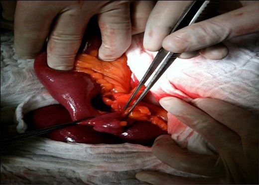
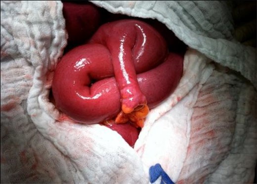
Following laparotomy, a gross distension of the small bowel and collapse of the large bowel was identified. The small bowel was subsequently delivered carefully and examined. Loops of distended small bowel were identified extending proximally from the duodenojejunal junction to the distal ileum. Thirty-five centimeters from the ileocecal valve, an 8-cm-long Meckel’s diverticulum had severe inflammation with the tip of it creating a symphysis band with the mesenterium (Fig. 1). Ileal loops were dilated at the superior part of the mechanical obstruction (Fig. 2). Obstruction was caused by trapping of a bowel loop by a mesenterium-diverticular band. After separating the band from the mesenterium, the ileal loop was released from the diverticulum. Resection of the Meckel’s diverticulum and closure of the bowel were done using a TA stapler. The small bowel was then decompressed and aspirated via the nasogastric tube. The patient recovered without any complications and was discharged after 7 days of hospitalization. The diverticulum was confirmed as Meckel’s diverticulum by histological examination.

35 centimeters from the ileocecal valve, an 8-cm-long Meckel’s diverticulum had severe inflammation with the tip of it creating a symphysis band with the mesenterium.

Ileal loops were dilated at the superior part of the mechanical obstruction.
DISCUSSION
Meckel’s diverticulum is the most common congenital malformation of gastrointestinal tract (most studies suggest an incidence of between 0.6 and 4%). The bulge is congenital (present at birth) and is a leftover of the umbilical cord (vitello-intestinal duct).
Gastrointestinal bleeding is a major cause of emergency hospital attendance in adults. Nearly 80% of this bleeding in adults originates proximal to the ligament of Treitz. The most common source of the lower gastrointestinal bleeding is colon, with less than 5% of bleeding from small intestine [1].
Meckel’s diverticulum is the most common cause of bleeding in the pediatric patients (due to the persistence of the proximal part of the congenital vitello-intestinal duct). It is a true diverticulum, typically located on anti-mesenteric border, and contains all three layers of intestinal wall with its separate blood supply from the vitelline artery. The mesenteric location of Meckel’s diverticulum has been documented [2]. In surgical textbooks, it is known by the rule of two: it is present in 2% of the population located at 2 feet (61 cm) from the ileo-caecal junction and is 2 inches (5 cm) long. Anatomical variations may exist. The average length of a Meckel’s diverticulum is 3 cm, with 90% ranging between 1 and 10 cm, and the longest being 100 cm.
The mean distance from the ileocecal valve seems to vary with age, and the average distance for children under 2 years of age is known to be 34 cm. For adults, the average distance of the Meckel’s diverticulum from the ileocecal valve is 67 cm.
Meckel’s diverticulum was first described in 1809 by the German anatomist, Johann Friedrich Meckel, the younger (1781–1833), who described it as a remnant of the omphalo-mesenteric duct [3]. This had been mentioned earlier by Fabricius Hildamus in 1598 and in 1671 by Lavater.
M.D. is lined with the typical ileal mucosa as it is in the adjacent small bowel. Ectopic gastric (20% according to recent data 4)—duodenal, colonic, pancreatic, Brunner’s glands, hepatobiliary tissue and endometrial mucosa may be found, usually near the tip [1] Complications are 3–4 times more frequent in males. The most common complications in adults are: obstruction due to intussusception or adhesive band (14–53%); ulceration (54%); diverticulitis and perforation. Its most common presentation is in children under the age of 2 years olds. Symptomatic Meckel’s diverticulum occurs in those aged 2–8 years. Carcinoid tumor, sarcoma, stromal tumors, carcinoma, adenocarcinoma, intraductal papillary mucinous adenoma of pancreatic tissue and vesicodiverticular fistulae are also rare complications [4, 5].
Obstruction can be caused by trapping of a bowel loop, by a volvulus of the diverticulum around a mesodiverticular band, a mesodiverticular band, intussusception as well as by an extension into a hernia sac (Littre’s hernia) [6]. Similarly, as in our case, obstruction can be caused by trapping of a bowel loop by a mesenterium-diverticular band. The important aspect of our case is a clear demonstration of the mesenterium-diverticular band from a Meckel’s diverticulum. High-resolution sonography usually shows a fluid-filled structure in the right lower quadrant having the appearance of a blind-ending, thick-walled loop of bowel [7]. Meckel’s diverticulum is difficult to distinguish from normal small bowel on CT. However, a blind-ending fluid or gas-filled structure in continuity with the small bowel may be revealed [6].
Abdominal CT is used for complicated cases such as intussusceptions and distinguish between lead point and non-lead point intussusceptions [8].
The preoperative diagnosis of Meckel’s diverticulum is still an outstanding challenge. Preoperative diagnosis of symptomatic Meckel’s diverticulum is difficult. This is particularly true in the patients presenting with the symptoms other than bleeding. In a study of 776 patients by Kusumoto et al., 88% of the patients presenting with bleeding had a correct preoperative diagnosis versus 11% with symptoms other than bleeding [9].
The question which could arise is what should a surgeon do when an asymptomatic Meckel’s diverticulum is discovered. A population-based study demonstrated a surgical complication rate of 7% for patients with a complicated Meckel’s diverticulum and a complication rate of only 2% over a 20-year period for incidental diverticulectomy. The main complication is being obstruction secondary to adhesions. The ability to remove the diverticulum safely is the deciding factor. Deaths associated with the removal of Meckel’s diverticulum have been reported. In our experience, we believe it is safe to remove an incidentally discovered Meckel’s diverticulum in the absence of any complicating conditions, such as diffuse inflammation. Contraindications to an incidental resection include the presence of ascites, contamination of the abdominal cavity by feces.
The treatment of choice for the symptomatic Meckel’s diverticulum is surgical resection. This can be achieved either by the diverticulectomy or by the segmental bowel resection and anastomosis, especially when there is palpable ectopic tissue at the diverticular-intestinal junction, intestinal ischaemia or perforation (bowel obstruction). Diverticulectomy is adequate for the incidental Meckel’s diverticulum or when diverticulitis presents at the tip of the diverticulum. Segmental resection is recommended when the base is inflamed or if the patient presents with melena. There have been suggestions that the morphologic characteristics of the diverticulum should be considered when deciding on the extent of resection, the risk of complications in short, broad-based diverticula compared with long, thin-based diverticula. Thin diverticula may increase the risk of volvulus, intussusception or torsion, whereas short, broad-based diverticula can predispose to trapping of an enterolith, leading to inflammation, hemorrhage or obstruction.
Clinical manifestations arise from complications of this true diverticulum that are most common in males under 40–50 years of age and with a diverticulum longer than 2 cm. Preoperative diagnosis of a complicated Meckel’s diverticulum may be challenging.
CONFLICT OF INTEREST STATEMENT
None declared.
FUNDING
None.


