-
PDF
- Split View
-
Views
-
Cite
Cite
Dania Badran, Safiyah Ismail, James Ashcroft, Pneumomediastinum following spontaneous vaginal delivery: report of a rare phenomenon, Journal of Surgical Case Reports, Volume 2020, Issue 6, June 2020, rjaa076, https://doi.org/10.1093/jscr/rjaa076
Close - Share Icon Share
Abstract
Pneumomediastinum is the presence of mediastinal air, which raises concern for life-threatening conditions such as esophageal perforation and mediastinitis. Here, we described the case of a young female with no previous past medical history, who developed spontaneous pneumomediastinum following uncomplicated spontaneous vaginal delivery (SVD) giving birth to a healthy newborn at full term. The incidence of benign pneumomediastinum following SVD is estimated at 1 in 100 000 deliveries. This case explores the etiology of this rare presentation, recommends essential investigations and advises on pertinent clinical considerations.
Introduction
Pneumomediastinum is defined as the presence of free air within the mediastinum. The estimated incidence of pneumomediastinum precipitated by increased intrathoracic pressure during childbirth and spontaneous vaginal delivery (SVD) is estimated to be one in every 100 000 deliveries [1]. The diagnosis of pneumomediastinum following SVD can be difficult when considering potentially life-threatening differentials such as tension pneumothorax, esophageal perforation and mediastinitis. We report the case of pneumomediastinum following SVD in a primigravid parturient giving birth to a healthy newborn at full term. This case explores the etiology of this rare presentation, recommends essential investigations and advises on pertinent clinical considerations.
Case Report
A young female primigravida patient with no previous past medical history underwent an uncomplicated SVD of a healthy newborn following spontaneous rupture of membranes at full term. The antenatal period was uneventful, and the patient delivered a healthy female newborn weighing 3.61 kg after 6 hours in labor with no analgesic requirement. Four hours following delivery, the patient alerted the on duty medical team to a swelling in the face and neck, which was noticeable bilaterally. Observations revealed blood pressure, heart rate, respiratory rate and oxygen saturation all within normal limits and the patient was afebrile. On physical examination, widespread crepitus was present across the right side of the neck radiating toward the jaw. Respiratory examination revealed no pathology with clear lungs on auscultation, equal bilateral air entry and no tracheal deviation.
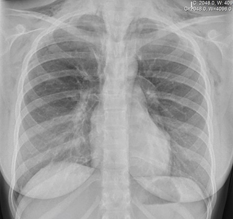
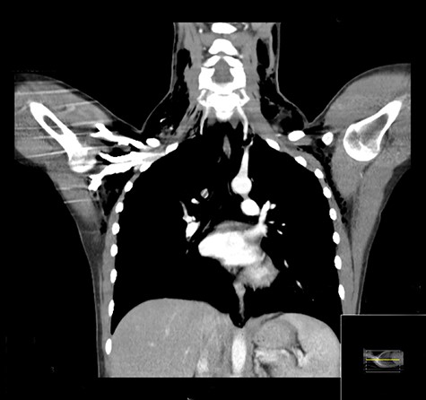
An urgent chest X-ray revealed extensive bilateral subcutaneous emphysema throughout the thorax and base of the neck with no visible pneumothorax or pleural effusion (Fig. 1). Thoracic and cervical computed tomography (CT) with contrast revealed a moderate pneumomediastinum and extensive subcutaneous emphysema throughout the neck, supraclavicular fossae, axillae and upper chest wall with no overt evidence of esophageal injury (Fig. 2).

Chest X-ray demonstrating pneumomediastinum with associated subcutaneous emphysema.

Coronal CT demonstrating extensive surgical emphysema in the neck, supraclavicular fossae, axillae and upper chest wall with moderate pneumomediastinum.
The patient was advised to remain nil-by-mouth for 5 days due to concern regarding an esophageal tear and further investigations including a water-soluble contrast swallow were carried out to exclude esophageal perforations. The esophagus was revealed to be intact, and there was no extravasation of oral contrast giving no evidence of an esophageal tear. In view of these results and improving symptoms, the patient was allowed to resume oral intake after 5 days. The patient underwent a repeat chest X-ray which confirmed the resolution of pneumomediastinum, and she was managed conservatively in hospital for a further 2 days (7 days post-partum in total) prior to discharge. Complete regression of symptoms and full recovery was noted on follow-up in the outpatient clinic 1 month later.
Discussion
Pneumomediastinum has a rare incidence which may be clinically undetectable and therefore underdiagnosed in the post-partum period [2]. The responsible pathophysiology relates to barotrauma, whereby the tearing of marginal alveoli into perivascular tissue planes traps air in the mediastinum [3]. This is associated with the Valsalva maneuver, which is widely used in the critical second stage of labor to direct pushing by raising intrathoracic pressure [4, 5]. However, high intrathoracic pressures combined with reduced vascular caliber create a pressure gradient, allowing air to dissect into the mediastinum along broncho-vascular sheaths [6]. This has been termed ‘Macklin’ effect and occurs in screaming, vomiting and forceful coughing, in addition to childbirth [7].
Patients presenting with pneumomediastinum precipitated by childbirth are of a younger age, primiparous, and have term or longer gestations [8]. Symptoms of pneumomediastinum in pregnancy may develop during labor but usually appear in the post-partum period. Common clinical signs and symptoms include chest pain, dyspnea, dysphagia, tachycardia and swelling of the face and neck. Such a clinical presentation combined with a chest radiograph revealing free air in the mediastinum is sufficiently diagnostic of pneumomediastinum in pregnancy [1, 2]. In the presence of atypical symptoms, for example vomiting or retching, a barium swallow test should be performed to identify the causes of secondary pneumomediastinum, such as Boerhaave’s syndrome, and a CT or endoscopy may be necessary in patients who are clinically deteriorating [8].
Following the correct identification of pneumomediastinum following SVD, patients should be managed conservatively with analgesia and rest [1, 2]. Previous guidance has suggested that Oxygen supplementation should be provided to this patient group to promote pneumomediastinum absorption into surrounding tissue by increasing nitrogen diffusion pressures in the interstitial space; however, further studies investigating oxygen as an adjunct to conservative management should be undertaken. The resolution of pneumomediastinum with oxygen supplementation has been noted to occur over extended periods of inpatient admission (up to 2 weeks) [9].
In this case, age, parity, clinical history and nature of symptoms in addition to chest radiograph images were all concordant with a diagnosis of pneumomediastinum following childbirth. However, further investigations due to concern for esophageal tear were undertaken, requiring restriction of oral food and drink which can have detrimental effects on the mother’s mental and physical well-being. In turn, there may be reduced ability to breastfeed, affecting the baby’s nutrition and early maternal bonding [10]. Furthermore, radiation exposure following CT scanning is a pertinent consideration in this young female patient, especially given its potentially unrequired role in diagnosis [1, 2].
This case demonstrates the importance of identification of pneumomediastinum in the obstetrics setting where its presentation may be vague and delayed. Its early detection can avoid distressing investigations and ensure an expeditious management, helping prevent nutritional deficits and prolonged hospital inpatient stays which may result in a reduced ability to breastfeed, ultimately affecting the newborn’s nutrition and early maternal bonding.
Conclusion
Pneumomediastinum is a self-limiting condition and a rare complication of childbirth. Current evidence suggests that pneumomediastinum following SVD can be treated conservatively with analgesics, rest and potential oxygen supplementation to enhance resolution. Diagnosis is based upon clinical history and positive chest radiograph findings. Awareness and consideration of this condition is important for the avoidance of prolonged nil-by-mouth protocols and unnecessary diagnostic investigations which can be potentially detrimental for both mother and child.
ACKNOWLEDGEMENTS
None to declare.
Conflict of interest statement
Authors have nothing to disclose.
FUNDING
Imperial College.
References
Author notes
Safiyah Ismail, Imperial College London South Kensington London SW7 2AZ Email: safiyah.ismail15@imperial.ac.uk
James Ashcroft, Department of Surgery & Cancer Imperial College London St Mary's Hospital Campus 10th Floor, QEQM Building Praed Street London W2 1NY Email: jamesashcroft36@gmail.com


