


Abstract
Cantrell syndrome (CS) is defined as congenital combination of five anomalies: defects at the lower part of the sternum, anterior diaphragm, midline supraumbilical abdominal wall, diaphragmatic pericardium and ectopia cordis. Antenatal screening should be performed to make an accurate prenatal diagnosis. The prognosis is usually poor with a high mortality early in life. The gold standard management is surgery but its prognosis remains poor. In many low-income settings prenatal examinations and surgery treatment are not possible. In the present case, we report a not surgery managed baby affected by CS, with good clinical conditions after 5 months.
INTRODUCTION
Cantrell syndrome (CS) or pentalogy is a rare congenital syndrome composed of defects of the abdominal wall, sternum, diaphragm, pericardium and congenital heart disease with an incidence ranging from 1: 65 000 to 1:200 000 live births with a male predominance of 1.35:1 [1, 2]. It can present with varying degrees of severity and different type of cardiac defect including ventricular septal defect, atrial septal defect, tetralogy of fallot, pulmonary stenosis and very mild sternal cleft to complete thoracoabdominal ectopia cordis [1]. Ectopic cordis (EC), a typical feature of CS, is defined as a partial or complete displacement of the fetal heart outside the thoracic cavity, and it is reported with a prevalence of 7.9:1 million live births [3].
Although the pathogenesis is still unclear, it seems that these anomalies are related to the developmental failure between 14 and 18 days of embryonic life involving an inappropriate differentiation of a segment of the lateral mesoderm [1]. Moreover, no genetic mutation has been yet discovered, although some cases pf CS have been associated with chromosomal abnormalities such as Trisomy 21, Trisomy 18 or Turner’s syndrome [1].
The prognosis is generally poor and depends on the severity of the cardiac anomaly, the defective skin area in terms of the susceptibility to infection and the available management [4, 5]. However, despite considerable advances in the management of CS, the survival rate is still fairly low and it depends primarily upon the type and severity of associated anomalies counting among the main causes of death respiratory and heart failure. This is particularly current in low-income settings where prenatal visits and ultrasonographic examination are usually not performed and surgery is not always available.
We report an extremely rare case of CS in a low-income setting treated with a conservative management.
CASE REPORT
A 27-year-old woman gravida 3 para 2 without the history of chronic diseases was referred to our hospital and delivered with a cesarian section, a 2.3 Kg female baby at 35 gestational weeks. No antenatal visit was performed and no maternal history of ingestion of unprescribed medicinal drugs, use of illicit pills, cigarette smoking or alcohol abuse was found.
The newborn clinically demonstrated the features of CS (Fig. 1), but no diagnostic studies were performed: computed tomography (CT) was not available, and no ultrasound specialist was present in order to perform one. In the country, there is no high-specialized public cardiac or thoracic surgery center and the family could not afford private referral. The only possible solution was to try a conservative management consisting on eosin dye and follow-up.
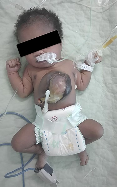
CS at birth.
After 6 weeks of treatment the newborn was discharge without infective and respiratory complications. After 5 months, the mother came for a clinical follow-up and the baby was in general good conditions with no bleeding or discharge from the site, no cyanosis, no other abnormalities, deformities or complications (Fig. 2).
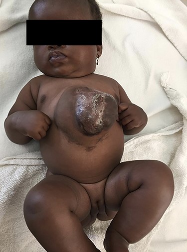
CS after 5 months of conservative treatment.
DISCUSSION
The approach to the CS should start during the prenatal period, when it is possible to make diagnosis simply by ultrasound starting from the first trimester due to the presence of the large omphalocele or the EC. A prenatal diagnosis gives parents the opportunity to be ready, take informed decisions or, sometimes, prenatal interventions [6]. Unfortunately, in our cases, as usually in low-income settings, no prenatal visit or test was performed. Indeed, the syndrome was diagnosed at the birth when visible and palpable pulsatile mass below the skin of the lower chest and upper abdomen was detected.
The postnatal evaluation should include both X-ray and ultrasound examination in order to reveal intra-cardiac anomalies, although computed tomography remain the most informative tools informing also on pericardium, diaphragm and abdominal viscera [7]. In our case, it was not possible to perform any test due to the lack of high-trained specialist and equipment. Again, also in term of management we were very limited. In fact, the best treatment is surgery with the aim of closing the chest wall defect either by primary chest wall closure or by using bone/cartilage as tissue graft or artificial prosthesis like acrylic plaques or Marlex mesh [8]. Of course, this procedure requires a multidisciplinary and highly specialized equipe and adequate equipment that, once again, are lacking at our hospital. For these reason, we proceeded just with a conservative approach trying to avoid infective complications. To date we have no solution for a cardiac surgery evaluation and intervention, although this case shows an extraordinary survival at 5 months. The most frustrating thing is that we are aware about the risk of the newborn to develop arrhythmia and low cardiac output that, unavoidably, will lead to the inability of the heart to adjust to the intra-thoracic pressure and consequently to death.
In conclusion, although CS remains a diagnostic and therapeutic challenge, especially in low income settings, this case highlights the urgent need to strengthen the antenatal care and counseling in low-income settings and to improve the quality of services for the management of diseases during the early days of life.
Conflict of interest statement
None declared
Funding
None.
Ethical statement
Written informed consent was obtained from the parents of the child for publication of this case report and any accompanying images.
