


Abstract
Wandering spleen is a rare condition characterized by the absence or underdevelopment of one or all spleen ligaments that fixate the spleen in the left upper quadrant. Many different terms refer to wandering spleen like dislocated spleen, ectopic spleen and displaced spleen. We report in this case a 13-year-old Syrian girl presented to the emergency department complaining of acute generalized abdominal pain with fever, anorexia and vomiting started 2 days prior to presentation. A splenectomy was performed, with uneventful postsurgical follow-up. Wandering spleen is prone to torsion and infarction resulting in acute abdomen and a life-threatening condition with high mortality rate reaching 50%. We advise the investigation of any recurrent episodes of chronic pain keeping up within mind this diagnosis.
INTRODUCTION
Wandering spleen is a rare case in which the spleen is hypermobile in the abdomen due to the absence or laxity of one or all spleen ligaments which fixate the spleen in the left upper quadrant; it may be congenital or acquired [1]. The condition occurs mainly in women in reproductive age, and its incidence is less than 0.2% [1, 2].
It may be asymptomatic, otherwise it may cause episodes of chronic abdominal pain or present as a mobile mass in the abdomen [3]. Due to the hypermobility of the spleen, it is highly prone to torsion causing a life-threatening acute abdomen condition [2].
The treatment of choice in wandering spleen is splenopexy, but when torsion and infarction occur, splenectomy is required [4].
This is a case of a 13-year-old female presented with acute abdomen and treated immediately with splenectomy; several investigations lead us to the diagnosis of infarction of wandering spleen.
CASE PRESENTATION
A 13-year-old girl presented to the emergency room with generalized, acute abdominal pain (worst in the hypogastric region) with fever 38.9°, anorexia and vomiting started 2 days prior to presentation.
The parents mentioned that they brought her 5 months ago with mild pain and abdominal mass; an ultrasonography showed no splenic tissue in upper left quadrant, which suggested a wandering spleen and a CT scan was ordered, but the parents ignored. She did not have history of trauma or any remarkable past medical history.
On examination, the abdomen was rigid with generalized tenderness and rebound tenderness. There was an evident mass bulging under the umbilicus. Heart rate was 123 bpm with otherwise normal cardiorespiratory function.
On admission, her hemoglobin was 11.3 g/dL, white cell count 19.6 × 10⁹/L, granulocytes 78.1% and CRP 21.6 mg/L. Her renal, liver function, amylase and lactate were within normal limits.
A Doppler ultrasonography showed that the spleen was in the hypogastric region with a diameter of 14 cm and a very low perfusion. The abdominal radiograph and the chest radiograph were normal.
She was prepared for an emergency laparotomy; the spleen was infarcted without any ligamentous attachments. The hilar vessels of the spleen were within a long mesentery. The splenic vessels were twisted three rounds around themselves. The other organs in the abdomen were normal (Fig. 1). A splenectomy was performed and the splenic vessels were ligated and then cut.
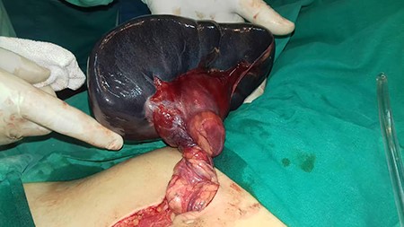
Infarcted spleen free of any ligamentous attachments and the twisted vessels.
Her recovery was uneventful, and she took vaccines dedicated for splenectomy patients.
DISCUSSION
Wandering spleen is a rare clinical condition that could be congenital or acquired caused by the absence or laxity of the spleen ligaments, which makes the spleen hypermobile in the abdomen [1]. Its incidence is less than 0.2% and represents less than 0.25% of total splenectomy procedures and occurs mainly in women in reproductive age [1, 2]. Approximately 500 cases have been reported, and children represent less than one-third of the cases [5].
Wandering spleen may present as a mobile mass, episodes of chronic pain or acute abdomen or could remain asymptomatic [3]. The hypermobile spleen is prone to torsion and infarction resulting in an acute abdomen and a life-threatening condition with a high mortality rate reaching 50%, which requires a delicate approach toward these patients [2].
Diagnosis can be challenging, and in the emergency setting, it is mostly diagnosed by CT, color Doppler ultrasonography and 99mTc-sulfur colloid [6].
Treatment of uncomplicated WS is splenopexy, which could be performed by creating pouches of peritoneum, retroperitoneum and omentum. Also, it could be performed by a synthetic bag which is then fixated to the diaphragm [5]. But when complications occur, splenectomy is the preferred procedure [4].
Our patient presented with an acute abdomen to the emergency department, and the spleen did not allow ultrasonography to view the structures behind, which made the diagnosis more challenging.
Finally, we advise the immediate investigation of any recurrent episodes of chronic pain, so that if there is such clinical entity, it could be diagnosed early and the spleen could be preserved as opposed to what happened with our patient.
CONCLUSION
Wandering spleen is an unusual medical case, and when it is complicated by a torsion and infarction, it turns into an abdominal emergency.
It is extremely helpful to use Doppler and CT for diagnosis; however, the diagnosis of wandering spleen can be challenging due to its nonspecific symptoms.
Finally, we advise the investigation of any recurrent episodes of chronic pain keeping up within mind this diagnosis.
ACKNOWLEDGMENTS
We would like to thank Joud Khalili, Joudi Sawas and Hamed Kozom (Medical students, Faculty of Medicine, University of Aleppo) for their support during this work.
Author’s contribution
Conception and design: BS and AA.
Analysis and interpretation of the data: BS, AA and EAA.
Drafting of the article: BS, AA, ARR, AM and RH.
Critical revision of the article for important intellectual content: KA and RA.
All authors read and approved the final version of the manuscript.
Consent for publication
An informed consent was signed by the patient.
Competing interests
We have no conflict of interest.
Funding
There are no funding sources.
