


Abstract
In this study, we report a unique case of aorto-bifemoral graft infection, which developed in a 47-year-old male patient after endovascular aortic aneurysmal repair (EVAR) and extra anatomic axillo-femoral bypass. The patient had previously been treated by EVAR for an infrarenal abdominal aortic aneurysm. Earlier, the EVAR was blocked by a thrombosis and treated with an extra-anatomic axillo femoral bypass, which then became occluded. The patient was then treated with an aorto-bifemoral bypass using a Dacron Y graft. A few months later, he was referred to our cardiovascular center with high body temperature, weight loss, inability to stand and walk, and very serious sepsis. A computed abdominal tomography scan revealed that a part of the graft proximal to the bifurcation had totally eroded into the proximal jejunum. We treated this patient with multiple surgeries, antibiotic administrations and hypochlorous acid irrigation without graft excision, which carries a high morbidity and mortality risks.
INTRODUCTION
Prosthetic graft infection after graft insertion for abdominal aortic aneurysm or occlusive vascular disease treatment is a rare complication with an incidence of 0.5–2.5%, which is associated with considerable high rate of morbidity and mortality, particularly when diagnosis is delayed [1]. The morbidity and mortality rate after this rare but serious complication has been estimated to range from 25% to 75% with an average of 45%. Inspite of the improvements in vascular surgery and graft technologies, the mortality and morbidity rate after abdominal aortic graft infection (AAGI) treatment has not decreased significantly [1–3].
Due to the nonspecific signs and symptoms, diagnosis may be difficult, particularly during the early stages of the disease. Extra-anatomic and in situ procedures are the widely used surgical revascularization methods of treatment with excision of the infected graft that may be followed by high morbidity and mortality [2–5].
CASE REPORT
A 47-year-old male patient was diagnosed with infra renal abdominal aortic aneurysm and treated with endovascular aortic graft repair (EVAR) in 2012. In October 2016, he presented with chest pain, fatigue, difficulty in breathing and lower limb pain. His angiography revealed nearly normal coronary arteries and occlusion of the EVAR. In October 2016, an axillo-femoral bypass was performed. In October 2017, he presented with left inguinal region discharge. A wound culture was obtained and revealed no micro-organism growth. Blood biochemistry revealed high level of C-reactive protein (CRP) of 47.8 mg/dl. The wound was cleaned and an empirical broad spectrum oral antibiotic was started. On 3 August 2017, the patient was admitted to our cardiovascular department because of claudication, pale lower extremities and abdominal pain. His examination revealed a positive pulse at the right femoral artery and no palpable pulse in the rest of the lower extremity arteries. A computed tomography (CT) angiography revealed axillo-femoral grafts occlusion. An aorto-bifemoral bypass was performed (21 August 2017). Post-operatively, the patient did fine and he was discharged on the seventh post-operative day. On 8 May 2018, the patient presented with both inguinal region discharge and fatigue. His CRP level was above the normal limits. Wound culture revealed no microorganism growth.
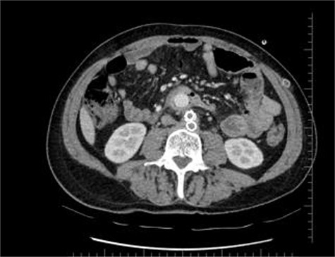
Axial section shows the two limbs of the EVAR, at the bottom. And on top of it, the Dacron straight graft part in the middle of the jejenum.
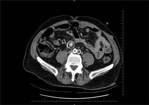
Distal CT axial section shows the two limbs of the EVAR, at the bottom. And on top of it, the Dacron straight graft part in the middle of the jejenum.
During the first days of December 2018, the patient visited another medical center because of fatigue, difficulty in breathing, lower back pain and inability to walk. His blood biochemistry revealed a very high CRP level (350 mg/dl), and complete blood count revealed a very high level of white blood cells (21 000/ul). An abdominal CT scan illustrated part of the graft was totally eroded into the small intestine (Figs 1–3). The patient was referred to my office and an infectious disease specialist. He had high body temperature (39.5°C) and was lying on the bed with lower extremities adducted to the chest. Antibiotic and supportive treatment was started. Emergency surgery was performed. We found an 8–10 cm of the straight part of the graft had become totally incorporated into the proximal part of the jejenum. The graft was freed, and 10–15 cm of the jejenum was resected and anastomosed. The area was irrigated with saline and hypochlorous acid. We then put a 20 x 10 composite mesh between the vascular graft and the intestine using a non-adhensive surface facing the intestine to prevent future graft and intestine interactions and erosion. After hemostasis, two drains were placed retroperitonally, and the abdomen was closed. On the fourth post-operative day, a high body temperature and high CRP levels were recorded. An abdominal CT revealed retroperitoneal abcess formation. During relaparotomy, the anastomosis was found to be intact, and the retroperitoneal abscess was drained and irrigated with saline. The drains were reinserted. After a month, he once again started to have high fever and high CRP levels. Again, an abdominal CT revealed abscess formation. This time, the abscess was approached transcutaneously. By making an incision in the proximal left lomber region, the abscess was drained. Another incision was made distally, and two drains were placed in the retroperitoneal area.
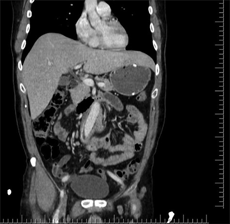
Coronal CT section shows straight part of the Dacron graft in the jejenum.
Patient treatment in addition to antibiotics, nutritional support and daily sludge irrigation with hypochlorous acid and saline for 3 months was undertaken. After 3.5 months, he was discharged.
DISCUSSION
Graft infection after surgery for abdominal aortic pathology (aneurysm or occlusion) is a rare complication associated with a high mortality rate. The mortality rate after this infection was found to range from 8% to 56% [6,7]. We think erosion of the graft into the jejunum tract occurred due to the continuous pressure of the underlying graft against that part of the small intestine lying on top of it.
Our case was unique in its development and treatment patterns. In general, AAGI cases are treated with graft excision and extra anatomic bypass or in situ repair [8]. However, our case had two grafts. One was the initially placed (EVAR) and on top of this one, the second Y Dacron aorto-bifemoral bypass was placed, which was achieved without excision of the occluded graft (EVAR). Normally, aortic graft infection treatment has a high risk of morbidity and mortality. It is not difficult to guess that this risk was even higher in our patient. Since he was diagnosed at a late stage, he was malnourished and quite septic. We tried a different treatment technique, which may have decreased the high risk of morbidity and mortality. To prevent future erosion of the graft into the intestine, the idea of using special kind of mesh (composite) with two surfaces, normal and non-adhesive ones, was attempted. This procedure was done by inserting a composite mesh under the mesentery with the non-adhensive surface facing the intestine mesenteric surface and the other one laid on top of the Dacron graft anterior surface. By doing this, we managed to separate the graft from the intestine and restrict the condition to the retroperitoneal area only. We could irrigate the area with saline and hypochlorous acid through the lodge drains. Using the combination of surgery (as described before), antibiotic treatment, and daily irrigation with saline and hypochlorous acid, we managed to eliminate the AAGI.
CONCLUSION
AAGIs can be treated by systemic antibiotics and surgical wound cleaning and irrigation with bacteriocidial solutions without graft resection. This procedure may lead to a decrease in the high mortality and morbidity rates, which are often seen in cases of classic AAGI treatment; however, more reports are needed to validate our findings.
