


Abstract
Inguinoscrotal hernia containing the urinary bladder is a rare entity found in 1–4% of inguinal hernias, while patients rarely present symptoms of urinary dysfunction. We present the case of a 79-year-old Caucasian male with acute renal dysfunction and incarcerated inguinoscrotal hernia containing the entire urinary bladder. The patient presented in the surgical emergency department due to an incarcerated right inguinoscrotal hernia and deteriorated renal function. Preoperatively, ultrasound imaging was performed, which showed the presence of the whole bladder in the hernia sac. The bladder was repositioned to its anatomic position and hernia was repaired through a modified Lichtenstein technique. In patients with inguinoscrotal hernia and acute urinary tract symptoms, surgeons should be aware of the possibility of inguinal bladder hernia. Preoperative imaging can help in preventing intraoperative bladder damage.
INTRODUCTION
Inguinal bladder hernia is a rare condition, occurring in 1–5% of inguinal hernias, and it was first described by Levine in 1951 [1]. It can be diagnosed during either preoperative or random imaging examination, or intraoperatively, where any portion of the bladder is identified in the hernia sac. Patients may be asymptomatic or complain about urinary dysfunction, such as double voiding, which may lead to the correct diagnosis. Renal failure, due to obstructive uropathy, and urinary infections can rarely complicate this condition.
We present the case of a patient suffering from incarcerated right inguinoscrotal hernia and acute renal dysfunction, in whom the presence of the entire bladder in the hernia sac was discovered preoperatively on ultrasound.
CASE REPORT
A 79-year-old Caucasian male patient presented to the surgical emergency department suffering from acute severe pain located in the right groin. The patient presented with fever (39.3°C) but did not mention diffuse abdominal pain, nausea or vomiting. His laboratory values included a white blood cell count of 11.470/μl (neutrophils: 80%) and C-reactive protein: 20.7. Renal function was acutely impaired (urea: 309 mg/dl, creatinine: 7.3 mg/dl), while previous laboratory results concerning renal function were normal. No urinary tract symptoms other than oliguria were mentioned.
On clinical examination, a sizable painful right-sided incarcerated inguinoscrotal hernia was found. All abdominal quadrants were otherwise soft on palpation, without specific signs of pain; bowel sounds were also normal.
Given the fact that the patient was still hemodynamically stable, and in order to identify preoperatively the content of the incarcerated hernia sac, prompt imaging with ultrasound was considered necessary. Imaging revealed the protrusion of the entire bladder through the right internal inguinal ring and extending into the scrotum (Fig. 1), as well as hydronephrosis of the right kidney (Fig. 2), justifying the patient’s renal dysfunction.
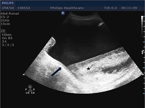
Longitudinal ultrasound image of the inguinal canal revealed a hernia that contained the entire urinary bladder. Notice the urothelium (arrow), as well as a great amount of debris (star).
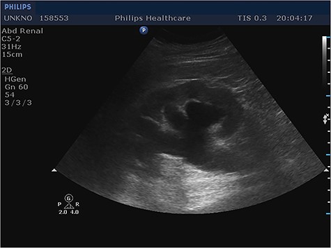
Longitudinal ultrasound image of right kidney showed hydronephrosis. Same findings noticed in contralateral kidney (not shown).
The patient was then submitted, under general anesthesia, to emergency surgery for hernia repair. During the operation, through a right inguinal incision, an indirect hernia sac was identified, which was filling the right hemiscrotum and contained the bladder and the balloon of the bladder catheter. The catheter balloon was temporarily evacuated and the bladder was inserted back in its anatomical place, since no signs of injury, necrosis or other pathology were present. Hernia repair was completed through modified Lichtenstein technique, with the use of a Parietex ProGrip mesh.
Postoperative period was uneventful, and renal function was normalized after 3 days; the patient was discharged from our surgical department on the seventh postoperative day. No renal dysfunction, surgical site infections, postoperative inguinal pain or recurrence were present during follow-up examinations.
DISCUSSION
Inguinoscrotal bladder herniation consists only 1–5% of inguinal hernias and occurs mainly after the fifth decade of age, with a morbidity of 1–4%, affecting males 10 times more than females. Other than male gender, it is suggested that factors such as obesity, weakening of the bladder tone and the abdominopelvic wall, and bladder-outlet obstruction with bladder distention are responsible for this condition [2]. Although small bladder hernias remain asymptomatic, patients with large bladder hernias usually complain about scrotal edema, decreased force of stream on urination, dysuria, double micturition through manual compression of the hernia and diminution of the scrotal edema after urination, called Mery’s sign [3]. Obstructive renal failure due to ureteric involvement is a rare finding. In our case, urologic history remained unknown, as the patient suffered from dementia and did not provide such details.
Preoperative ultrasonography, computed tomography (CT) scan, magnetic resonance imaging and cystography are considered effective imaging techniques and can decrease the rate of intraoperative bladder damage. Ultrasound is the most accessible and cost-effective method, showing a hypoechoic mass protruding from the bladder to the scrotum through the inguinal canal. Voiding cystography is considered the best imaging technique, demonstrating a dog-ear shaped bladder in the scrotum; cystoscopy can be used for the evaluation of the prostate and in cases of gross hematuria [4]. CT scan is indicated in cases of obesity, males more than 50 years of age and presence of lower urinary tract symptoms. In case of bilateral bladder hernia, ‘pelvic micky mouse sign’ is demonstrated in CT scan.
When not diagnosed preoperatively, bladder hernia is identified either intraoperatively, usually due to a concomitant iatrogenic injury, in about 12% of bladder hernias, or postoperatively, due to complications [5]. In case of intraoperative identification of the injury, however, catheterization and immediate repair of the injury are feasible. Further injury or ligation of the ureter should be avoided, as it can lead to threatening postoperative complications, such as sepsis, massive hematuria and fistula formation. Hernia neck’s diameter less than 5 mm, bladder wall necrosis, perforation and presence of a diverticulum or a bladder tumor are indications of partial bladder resection [6]. In these cases, a urologist should be consulted. In our case, a urologist was also consulted and no further intervention was suggested as needed.
When resection is not needed, the protruding bladder can be reduced to its anatomic place; hernia repair is then performed through the Lichtenstein tension-free repair, as in our patient, or through the open extraperitoneal technique. Laparoscopic approaches have also been reported. There is still no consensus on the ideal repair technique; surgical approach depends on the surgeon’s preference and the patient’s condition. Nonetheless, careful identification of every anatomic structure inside the hernia sac is important in order to avoid any iatrogenic injury [7, 8].
In conclusion, the presence of the entire bladder in the inguinoscrotal hernia is a rare finding that often remains unrecognized until surgical intervention. In cases of high suspicion, preoperative imaging is crucial in order to avoid intraoperative iatrogenic damage of the bladder.
CONFLICT OF INTEREST STATEMENT
The authors have no conflicts of interest to declare relevant to this article.
FUNDING
None.
