-
PDF
- Split View
-
Views
-
Cite
Cite
Shaurya Jhamb, Harshil Pillai, Christopher Maguire, Pranavan Palamuthusingam, Incidental finding of heterotopic supradiaphragmatic liver, Journal of Surgical Case Reports, Volume 2020, Issue 10, October 2020, rjaa394, https://doi.org/10.1093/jscr/rjaa394
Close - Share Icon Share
Abstract
Here we describe a case of heterotopic, supradiaphragmatic liver in a 65-year-old woman who was referred for investigation of a soft tissue gallbladder mass. Contrast-enhanced magnetic resonance imaging revealed adenomyomatosis of the gallbladder and supradiaphragmatic accessory liver tissue. This is a remarkably rare normal variant.
INTRODUCTION
Heterotopic, supradiaphragmatic liver tissue is a rare phenomenon. Since 1957, only 28 cases have been reported in the literature [1]. Clinically, it presents a diagnostic challenge as most patients are asymptomatic and it is often discovered incidentally [2]. Here, we present a case of intrathoracic liver diagnosed on contrast-enhanced magnetic resonance imaging (MRI) in a patient with no significant previous surgical history or trauma.
CASE REPORT
A 65-year-old Caucasian woman was referred to our hospital for further workup of a soft tissue gallbladder mass. The patient initially presented to their primary care physician with a history of postprandial, non-specific epigastric pain. She denied any associated symptoms or history of trauma and clinical examination was unremarkable. Her past medical history included hypertension, thoracic spine arthritis and hypercholestrolaemia. A subsequent ultrasound scan (USS) showed an irregular thickening of the anterior wall of the gallbladder measuring 12 × 9 × 20mm. Contrast-enhanced computed tomography (CT) of the abdomen revealed ill-defined, lobulated soft tissue masses within the gallbladder.
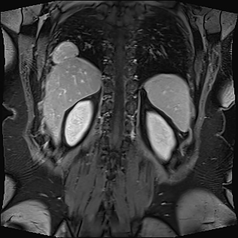
Given the ambiguous morphology of the lesions and the inconclusive CT and USS findings, an abdominal MRI was arranged. The MRI showed a distended gallbladder with multiple calculi in the lumen measuring 2–3 mm. In addition, segmental focal concentric thickening consistent with adenomyomatosis was identified at the body and fundus of the gallbladder. Notably, a multilobulated solid lesion measuring 5.7 × 2.9 × 3.6 cm was described in the right hemithorax, closely abutting the right costophrenic recess (Fig. 1). The mass appeared to be continuous with the right hepatic lobe via a narrow pedicle and demonstrated similar architecture and vessel continuity with the liver.

Multilobulated solid lesion measuring 5.7 × 2.9 × 3.6 cm seen in the right hemithorax adjacent to a right costophrenic recess, closely abutting the liver and appears to be continuous with the liver parenchyma with a narrow pedicle/area of contact.
The patient was diagnosed with uncomplicated cholelithiasis with an incidental finding of supradiaphragmatic liver noted as being an exceedingly rare normal variant. A follow-up USS in 4–6 months was organized to confirm no interval progression of appearances.
DISCUSSION
This case discusses the presence of an intrathoracic, accessory liver lobe—an extremely rare entity. Two primary aetiological mechanisms have been proposed in the literature [3]. In patients with a transdiaphragmatic pedicle connecting the accessory lobe to the orthotopic liver, an embryological origin for the defect has been discussed [1, 3]. The development of the liver and diaphragm are closely related. The hepatic bud extends ventrally from the caudal segment of the foregut and in the fourth week of gestation, intercalates into the adjacent mesenchyme of the septum transversum [4]. Concomitantly, the central tendon of the diaphragm develops from the septum transversum and fuses with the peripheral pleuroperitoneal membranes, thereby separating the abdominal and thoracic cavities [5]. It is possible for a section of proliferating hepatic tissue to penetrate the thoracic cavity prior to the closure of the diaphragmatic membrane leading to pulmonary sequestration of hepatic tissue [1, 3, 6]. It is also possible for an independent hepatic bud to form in the thorax, leading to the pathogenesis of a supradiaphragmatic, ectopic liver without any connection to the orthotopic liver [1, 3].
The other mechanism hypothesised is the herniation of liver parenchyma into the thoracic cavity in patients with a prior history of surgical or high-impact trauma [3, 7]. These cases primarily occurred in older patients and often presented as a newly identified supradiaphragmatic mass on imaging studies.
The intrathoracic accessory liver lobe poses a diagnostic challenge for clinicians. Patients are frequently asymptomatic or present with non-specific symptoms such as dyspnoea, cough and chest pain [1, 8]. Furthermore, most imaging modalities often result in equivocal findings. X-rays only reveal a radio-opaque thoracic mass [9] and while contrast-enhanced CT scans are useful in elucidating the relationship of the mass to surrounding structures, they are unable to provide a precise diagnosis [4, 8]. However, contrast-enhanced MRI has emerged as an effective tool in differentiating supradiaphragmatic liver from other potential diagnoses. On T2 weighted sequences, the ectopic tissue demonstrates similar signal intensity to the liver parenchyma and on contrast-enhanced T1-weighted images, the tissue exhibits equal enhancement when compared to liver parenchyma [1]. Furthermore, as presented in this case, the presence of a transdiaphragmatic pedicle connecting the mass to the liver, as well as evidence of vessel continuity with branches of the hepatic and portal veins further aids the diagnosis of a supradiaphragmatic accessory liver lobe.
Despite its exceptional rarity, supradiaphragmatic liver should enter the differential diagnosis for an intrathoracic mass with similar signal to orthotopic liver—particularly when newly identified in patients with a history of trauma. The vast majority of patients are asymptomatic and require no surgical intervention; however, these ectopic masses should be followed up to ensure there is no interval progression or development of disease.
CONFLICT OF INTEREST STATEMENT
None declared.


