-
PDF
- Split View
-
Views
-
Cite
Cite
Ammar Omran, Mhmmad Nassif, Nabila Salhab, Aras Abdo, Mohammad Ahmad Almahmod Alkhalil, Norma Taishori, Adnan Dayoub, Early separation of omphalopagus conjoined twins: a case report from Syria, Journal of Surgical Case Reports, Volume 2020, Issue 1, January 2020, rjz374, https://doi.org/10.1093/jscr/rjz374
Close - Share Icon Share
Abstract
Omphalopagus twins are one of many forms of conjoined twins sharing part of the gastrointestinal system and abdominal wall. This type of twins has the best chance of survival if successfully separated. Surgical approaches in these cases are generally preferably elective, but sometimes separation may be urgently needed due to life-threatening complications, such as hemodynamic instability, death of either twin, necrotizing enterocolitis, among many others. We report a case of successfully separated omphalopagus twins at day two of life.
INTRODUCTION
Conjoined twins are a rare phenomenon of a monochorionic monoamniotic twin. The incidence varies from 1 in 50 000 to 1 in 10 0000 live births [1]. Five types of conjoint twins are commonly identified: thoracopagus, omphalopagus, pygopagus, ischiopagus and craniopagus. Omphalopagus is the least recurring with an incidence of 0.5%. Termination of pregnancy may be offered as a course of action if diagnosis determines a case of conjoint twins following a prenatal ultrasound; however, in underdeveloped countries, owing to the lack of adequate maternal care facilities, this is likely to be delayed.
Here, we describe a case of omphalopagus twins referred to our institute without antenatal diagnosis and preparation. The present case report is unique in the sense that it not only reports a rare condition but also highlights the fact that, notwithstanding medical advances, early separation of twins may help to manage necrotizing enterocolitis (NEC) when it occurs or may contribute to avoiding its occurrence altogether.
CASE REPORT
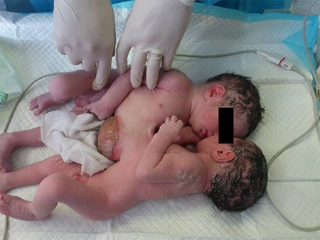
A 34-year-old mother gave birth to male omphalopagus twins via caesarian delivery (Fig. 1). The combined birth weight of the babies was 4.5 kg. The babies were designated as ‘twin R’ and ‘twin L’ (right and left), and were fused from epigastrium to umbilicus and had a single umbilical cord. The fusion area was covered with skin superiorly and a membrane inferiorly in the omphalocele. The left twin had two episodes of vital signs deterioration and cyanosis for few minutes in the neonatal intensive care unit (NICU). Both babies cried immediately after birth and passed urine and meconium.

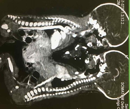
Ultrasonography of abdomen revealed fusion of liver but separate organ systems. A contrast computed tomography (CT) scan showed a large fusion in livers with secondary vascular connections, and twin L’s vena cava was pulled by the conjoined liver anteriorly (Fig. 2). Other organs were not shared. A 2D echo study of twin R revealed small Patent ductus arteriosus (PDA), while twin L had small atrial septal defect (ASD) with PDA. Blood investigations revealed haemoglobin (Hb) of 14.7 g/dl for Twin R and 13.5 g/dl for Twin L, with normal biochemistry values for both.

A contrast CT scan showing differential enhancement of livers of both twins and pulled vena cava for left twin.
Written informed consent was obtained from the parents, specifying saving the baby with the better chance of survival should conditions arise where such a decision became necessary.
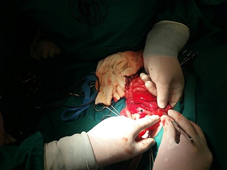
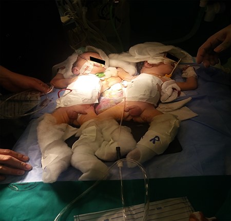
On the second day after birth, the decision to surgically separate was taken due to the risk of membranous part rupture and the pulled vena cava, which led to unstable hemodynamic in the babies. An extrahepatic biliary system of both twins was found with two gall bladders, but there was a fusion of 5 cm in the parenchyma (Fig. 3). First, the liver fusion was separated and the large abnormal vein was detected and ligated, and then the intestine was connected with a duct in the location of Meckel diverticulum and malrotated. The connection was cut and resutured and malrotation was corrected. After that, the lower part of sternum was connected with cartilaginous bridge, and many secondary vascular connections were ligated. Drainage for each twin was performed, and so was the primary closure of abdominal defect using the conjoined skin and fascia. Following a 5-h surgery, we had two complete babies with normal vital signs and good cosmetic results (Fig. 4). The need for ventilation support was indicated postoperatively. Twin R extubated after 24 h, but twin L, with the pulled inferior vena cava, had NEC. This was managed conservatively, but its condition dramatically deteriorated and he died on day 10 after the surgery. The other baby, however, did well and was discharged from NICU after 35 days. Five months later, the baby had bilateral herniotomy and he is doing well now.

Intraoperative view: connecting part of liver between twins, asterisk represents right twin’s gall bladder.

DISCUSSION
Each surgery of conjoined twins has its unique circumstances, and its success depends on points of attachment, organs shared and, definitely, the experience and skill of the surgical team. All these factors make the surgery range from possible to impossible.
First of all, antenatal diagnosis is very important in the management of conjoined twins [2], but, unfortunately, in the case under study, ultrasonography was possible but the face-to-face position of the twins made it hard to detect the fusion antenatal. Thus, the case was referred to our center without any planning done before delivery. Although females in conjoined twins predominate at the ratio of 3:1 [1, 3], our case involved male conjoined omphalopagus twins. The timing of separation is probably best to plan on an elective basis, but sometimes emergency separation may be needed. It is controversial, however, since delaying for a few months might increase the chances of survival [4]. Early separation is indicated when one twin threatens the life of the other [4]. Although major cardiac anomalies are contraindications of separation, variations in cardiac functions have been observed, and, in our case, the hearts were normal.
Conjoined biliary tract is reported in 25% of cases of omphalopagus twins [5]. The routine evaluation of cross-circulation is performed using many methods, such as Tc-99 m micro colloidal human serum albumin, hepatobiliary iminodiacetic acid (HIDA) scan [6]. Another possible method of evaluating cross-circulation is the contrast CT scan [4], and intravenous fluorescein can be used to demarcate large liver juncture [7]. However, the contrast CT scan did not provide us with sufficient information about biliary tracts, while the other investigations were not available at our center. Thus, it was only intraoperatively that we were able to detect separate biliary systems and two gall bladders.
NEC has been reported as a common complication in conjoined twins not only before [8, 9] but also after separation [10]. The postoperative period was uneventful for the right twin, whereas the left twin had NEC and died 10 days after the surgery. Accordingly, it is not only the shape or region of fusion that determines prognosis for a conjoined twins’ surgery, but also the physiological effects resulting from such fusion. The separation we performed was successful, and it was the first reported successful surgery to separate conjoined twins in our country in spite of the many unfavorable and challenging circumstances the country has been facing.
ACKNOWLEDGEMENTS
None.
CONFLICT OF INTEREST STATEMENT
None declared.
Funding
The authors did not receive any funding to conduct this study.
Ethical Approval
No ethical approval was needed.
Consent
Written informed consent has been duly obtained.


