-
PDF
- Split View
-
Views
-
Cite
Cite
Ammar Omran, Bardisan Sleman Gawrieh, Aras Abdo, Mohammad Ali Deeb, Mohammad Almahmod Khalil, Waseem Shater, Amyand hernia: scrotal pyocele, associated with perforated vermiform appendix and complicated by testicular ischemia in neonate, Journal of Surgical Case Reports, Volume 2019, Issue 9, September 2019, rjz265, https://doi.org/10.1093/jscr/rjz265
Close - Share Icon Share
Abstract
The presence of vermiform appendix in an inguinal hernia sac is known as Amyand’s hernia. This research paper examines the case of a 28-day-old Syrian male presented with a history of an infected right-sided hydrocele from the age of 14 days. Upon admission, ultrasonography was reported as a right testicular torsion. Accordingly, emergency surgical exploration was performed, and by exposing the spermatic cord fascia, 7 mL of pus was drained, revealing the cecum and perforated appendix lying beside the right testis, which showed evidence of ischemia and bluish discoloration.
Introduction
The presence of the vermiform appendix within an inguinal hernia was first described by Claudius Amyand in 1736. Since then, the term Amyand’s hernia has been in use to describe an appendix in the hernia sac whether the vermiform appendix is normal, inflamed or perforated or not [1]. It has an incidence of 1% and is complicated by acute appendicitis in 0.08% of cases [2]. Clinical presentation varies, depending on the extent of inflammation of the appendix. Pediatric patients may present with nonspecific symptoms, such as irritability and lethargy, along with a tender mass near the inguinal canal [3].


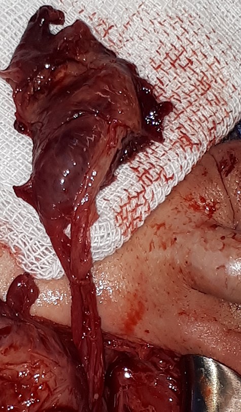
The right testicle showed evidence of ischemia and bluish discoloration.

Case Report
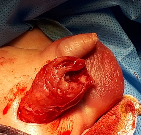
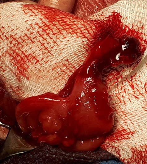
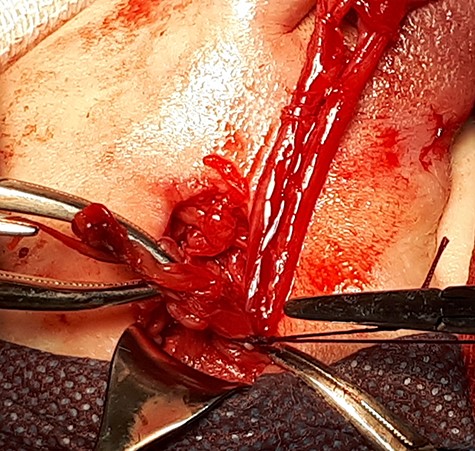
A 28-day-old Syrian male with a history of infected right-sided hydrocele and spermatic cord edema from the age of 14 days, as an ultrasonography report showed, was admitted to our Emergency Department. The right hemiscrotum swelling had become larger and erythematous 5 days prior to presentation. On admission, the patient was febrile (39°C) but neither vomiting nor suffering from bowel habit disorders. Objective local examination showed irreducible, rigid, large inguinal scrotal swelling covered by red edematous skin. Inflammatory markers were as follows: C-reactive protein, 150 mg/L; leukocytosis, 22.5 × 10⁹/L. Abdominal X-rays revealed no pathological elements. Ultrasonography was reported as right testicular torsion with an unclear fluid collection and gas detected around the right testis, spermatic cord oedema and the hernia sac; however, blood flow to the right testis could not be verified by the physician. Emergency surgical exploration was performed through an incision made on skin crease lateral to the pubic tubercle. An edematous, thickened spermatic cord fascia was observed (Fig. 1), and the fascia of the right testis was extracted out of the incision using blunt dissection. By exposing the spermatic cord fascia, 7 mL of pus was drained, revealing the cecum and perforated appendix, especially on its distal part, lying beside the right testis (Fig. 2). The testicle showed evidence of ischemia and bluish discoloration (Fig. 3). An appendectomy was performed, and the cecum was redelivered into the abdomen via the hernia sac, which underwent high ligation and excision (Fig. 4). The testicle looked viable and discoloration was corrected before closing. Orchiopexy was performed by anchoring the right testis to the scrotal wall. The patient was given a course of ceftriaxone and metronidazole. The postoperative course was uneventful, and the patient was discharged home after 5 days. After 1 month, the patient underwent a scheduled left-side herniotomy. Follow-up was maintained up to 6 month following the first surgery, and no complications or recurrence was observed.
Discussion
The presence of appendix in an inguinal hernia sac, known as Amyand’s hernia, is usually diagnosed intraoperatively. The signs and symptoms may mimic inguinal lymphadenitis, epididymo-orchitis, hydrocele of the spermatic cord or testicular torsion, and it is mostly delayed in diagnosis and treatment [4, 5]. In our case, the patient had a history of infected hydrocele and was misdiagnosed with testicular torsion by ultrasonography. The pathophysiology of Amyand’s hernia and its relationship with appendicitis are unknown. Some authors consider it an accidental finding; for others, a decrease in vascularization during incarceration and the maneuver to reduce the hernia result in inflammation of the appendix [6]. Our patient did not undergo any maneuver to reduce the hernia. It has been noted that both CT scans and ultrasonography can be helpful in providing a preoperative diagnosis [6, 7]. In our patient’s case, we had an ultrasound, which indicated testicular torsion and pyocele. However, an inflamed appendix was not initially suspected. Due to the low incidence of appendicitis but high incidence of incarcerated inguinal hernia in infants, the diagnosis of appendicitis in the hernia sac is delayed, similar to the case under examination, and thus the risk of perforation is higher. The classical treatment of Amyand’s hernia includes appendectomy and hernioplasty via the same incision; however, the need for prophylactic appendectomy during repair of Amyand’s hernia is subject to debate [1]. Laparoscopic surgery has become a more popular approach, is both diagnostic and therapeutic and may have benefits in some cases [3]. Since the first report by Waldbaum and Green, many cases of infected hydroceles in neonates have been described in the literature. An infected hydrocele in a neonate may be confused with testicular torsion, incarcerated inguinal hernia or epididymo-orchitis [8–10]. The cause of infected hydrocele remains to be defined. We report a case of a neonate with a history of infected hydrocele diagnosed with Amyand hernia, which led to testicular ischemia owing to delayed diagnosis. As in our case, accurate diagnosis before exploration is difficult in both infected hydrocele and Amyand hernia [4, 8]. In ultrasonography, an infected hydrocele usually appears as hyperemia of the testis and epididymis in combination with the presence of a surrounding heterogeneous fluid collection [9]. In our case, the ultrasonography was conducted as right testicular torsion and there were detected unclear fluid collection with gas around the right testis, spermatic cord oedema and inguinal hernia sac. Blood flow to the right testis was not visible. Cases of peritonitis leading to pyocele formation have shown infection with Escherichia coli [10]. In the case under examination, cultures of the pyocele fluid grew E. coli. To our knowledge, this is the first reported case describing infected hydrocele and Amyand’s hernia as associated entities. Surgical drainage is considered the definitive therapy of pediatric pyoceles. Many authors report cases of pediatric pyocele, where treatment was with intravenous antibiotics alone [9, 10].
Conclusion
Amyand hernia should be considered in neonates with infected hydrocele. Surgical exploration and appendectomy with inguinal herniotomy along with IV antibiotics is the treatment of choice.
Conflict of interest statement
None declared.


