-
PDF
- Split View
-
Views
-
Cite
Cite
Ahmed Nazer, Ahmed Al Awwad, Ahmed Aljuhayman, Ahmed Alasker, Saeed Bin Hamri, Stone fragments migration into subcapsular renal hematoma post flexible ureterorenoscopy (unique presentation), Journal of Surgical Case Reports, Volume 2019, Issue 5, May 2019, rjz125, https://doi.org/10.1093/jscr/rjz125
Close - Share Icon Share
Abstract
This is a case report of 45-year-old female patient who presented with right flank pain. Abdominopelvic CT scan showed right renal pelvis stone measuring 20 mm. Right flexible ureterorenoscopy with holmium laser was performed in our institute. Postoperatively, she was febrile and pale. Immediate Abdominopelvic CT scan was obtained which revealed a large right subcapular hematoma. Conservative management was maintained for a week. Two months later, repeated Abdominopelvic CT scan showed regression of right subcapsular renal hematoma with stone fragments migration into the perinephric space as a first presentation in the world.
INTRODUCTION
Stone disease is a common pathology in Saudi Arabia. Advances in endourology and holmium laser permitted urologists to expand their stone management modalities. Flexible ureterorenoscopy with holmium laser (FURS-L) is one of the new modalities in treating renal stones with low morbidity. Nevertheless, subcapsular hematoma is considered as a rare complication post FURS-L. Out of 250 interventions were performed in our center, we reported this unique presentation of stone fragments migration into a subcapsular renal hematoma.
CASE REPORT
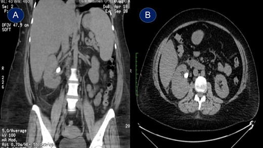
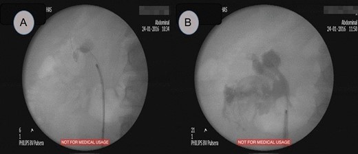
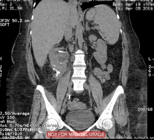
This is a 45-year-old female patient known to have diabetes mellitus and old cerebrovascular attack presented with right flank pain. Laboratory investigations revealed normal WBC count with a hemoglobin 10.4 mg/l. Radiological investigation showed right a 20 mm right renal stone (Fig. 1). Patient underwent FURS-L using a 10/12Fr Ureteral Access sheath. We did endoscopic renal exploration plus laser lithotripsy using Flex-Xc STORZ. The irrigation was under hydrostatic pressure of 80 cm H2O. The procedure was uneventful with an operative duration of 88 minutes. However, severe extravasation was noted at the end (Fig. 2). Six hours postoperatively the patient started to have high grade fever with a sudden drop of hemoglobin level to 6.6 mg/l. Immediate abdominopelvic CT scan with contrast was carried out showing severe right subcapsular renal hematoma. This complication was managed conservatively through proper antibiotics, blood transfusion and good hydration for 7 days. The patient was seen in the outpatient clinic 2 months later with a new abdominopelvic CT scan which showed a regression of subcapsular renal hematoma and surprisingly migration of stone fragments into the regressed subcapsular hematoma (Fig. 3). On the other hand, the upper urinary tract was free of stones.

Plain abdominopelvic CT scan, coronal view (A) and axial view (B) showing right renal pelvic stone measuring 20 mm.

Fluoroscopic study, (A) showed a radiopaque shadow at level of L2, (B) showed extravasation of contrast at the end of surgery.

Plain abdominopelvic CT, coronal view showed migrated stone fragments into the regressed right subcapsular renal hematoma.
DISCUSSION
Subcapsular renal hematoma (SRH) is an uncommon complication and it is rarely described after FURS-L. The first detailed case was reported by Bansal et al. [1] Bai and colleagues summarized 2848 patients post FURS-L, SRH was reported in 11 patients (0.4%) [2]. Out of 11 patients three resolved completely without intervention, six were managed with a percutaneous drainage, while two underwent open surgery with lumber incision. Chiu and colleagues reported a similar incidence of SRH post FURS-L [3]. In the series, they reported 4 (0.36%) SRH after 1114 FURS-L. Risk factors of SRH mentioned by Bai and co-workers include larger stone size (1.4 vs 0.9 cm), severe ipsilateral hydronephrosis, longer operation duration (41 vs 33 min), and higher perfusion pressure of hydraulic irrigation (176.8 vs 170.2 mm Hg) [2]. We presented our case out of 250 patients underwent FURS-L, this represented 0.4%, which is comparable to previous studies [1–3]. In our case, conservative management was sufficient. However, as mentioned by Chiu and colleagues the management of SRH should be customized for each patient. As far as we know, our case report is the first complication in the world where stone fragments migrated into perinephric space. Although we had no obvious trauma to pelvicalyceal system and the operative procedure was uneventful the most probable etiology of this incidence is due to forniceal rupture caused by high pressure or guidewire manipulation aggravated by longer operative time as mentioned by Bansal and colleagues [1].
CONFLICT OF INTEREST STATEMENT
None declared.


