


Abstract
Extracardiac adult rhabdomyoma is a rare benign tumor, which mainly occurs in the head and neck region and originates from striated muscle tissue. We report a 64-year-old male with simultaneous diagnosis of three adult rhabdomyomas including the soft palate and performed a review the literature on multifocal adult rhabdomyoma (mARM). Including the present case, 27 mARM with a range of 2–7 lesions per patient were collected. Mean age at diagnosis was 65 years with a male (23) to female (4) ratio of 5.75:1. Common localizations were parapharyngeal space (35%), larynx (14%), submandibular (13%), paratracheal region (14%), tongue (10%), floor of mouth (9%), neck (3%) and soft palate (2%). In accordance to this review, this the first case of mARM with involvement of the soft palate.
INTRODUCTION
Rhabdomyomas (RM) are amongst the rarest benign tumors in humans. They originate from striated muscles and are classified in cardiac (CR) and extracardiac rhabdomyomas (ER). The cardiac type is commonly associated with genetic abnormalities and appears almost solely in the hearts of infants. In dependence of the skeletal muscle differentiation, ER can be subdivided into a rare fetal (FRM), a more common adult form (ARM)—with preferred occurrence in the head and neck area—and a genital form (GRM) which appears in the vulva and vagina of women. About 3/4 of ER are located in the head and neck area awhile just 14% are found in the genital region [1]. A multifocal adult rhabdomyoma (mARM) is even a more rare tumor. We hereby report the first case of mARM involving the soft palate.
CASE REPORT
A 65-year-old male was referred to our department with a constant urge to clear the throat for a time span of 12 months. In addition, the patient stated his progressing inability to swallow food. Due to these symptoms, a resection of the thyroid gland had been carried out earlier showing struma colloides nodosae as well as bilateral parathyroidal adult rhabdomyomae. Even so, there was no relief in symptoms. At outpatient presentation at his dentist, a slight swelling of the soft palate was felt and the patient was referred for further therapy. Endoscopic examination as well as magnetic resonance imaging (MRI; Fig. 1) unveiled a tumor on the right side of the soft palate with a size of 5 × 5 cm2 and distinct demarcation to the surrounding tissue. Subsequently, the lesion was completely excised (Fig. 2) and histopathological analysis was conducted that showed a circumscribed but not encapsulated mesenchymal tumor with polygonal cell formation. The cells presented a granular cross-striated eosinophilic cytoplasm, large round vesicular nuclei and so called spiderweb cells (Fig. 3). Immunohistochemically, the cytoplasm of the cells was 100% positive for antibodies to desmin and S100 (Fig. 4). Additional immunohistochemical markers showed slight nuclear positivity for myogenin and nuclear negativity for AE1/3, CD68 as well as melan A. The histological examination confirmed ARM without signs of malignancy. At a total follow-up of 3 years, including MRI scan, no signs of recurrence were detected.
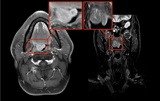
MRI scans of ARM involving the soft palate.
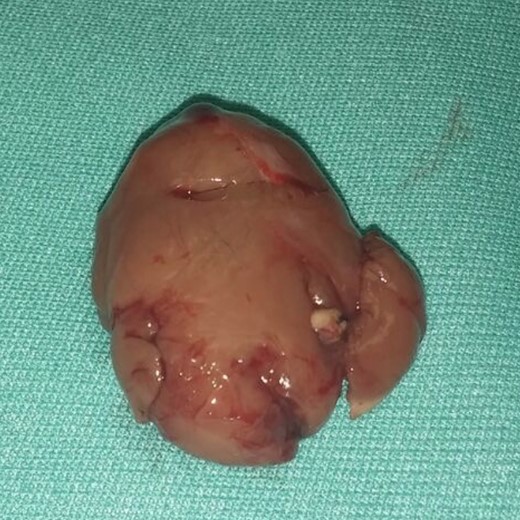
ARM of soft palate after excision.
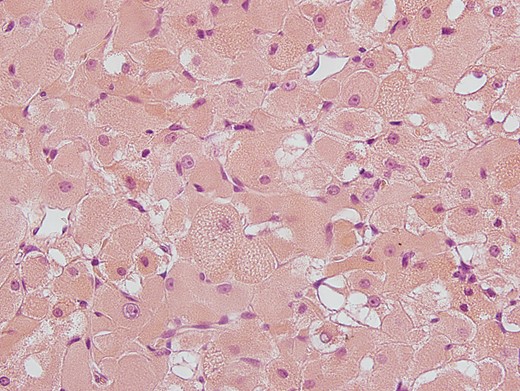
Hematoxylin-eosin (H&E) staining: polygonal cell formation with granular cross-striated eosinophil cytoplasm and large round vesicular nulei are seen.
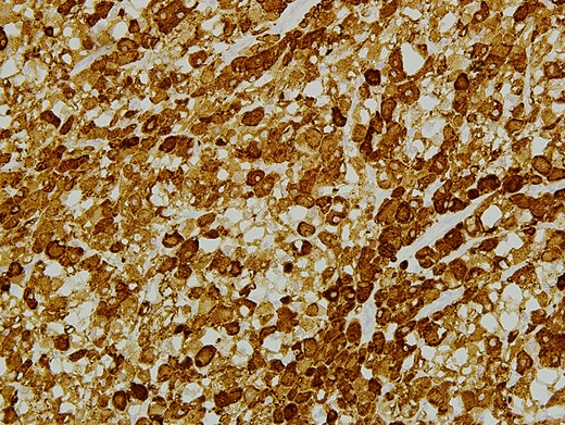
Immunohistological staining of EARM with desmin antibodies. Cytoplasma with positive antibodies for desmin is seen.
DISCUSSION AND REVIEW OF LITERATURE
RM have been first described by Weber [2]. The rare benign tumor originates of striated muscle cells with varying degrees of differentiation and maturity. It can be divided into cardiac and ER. They are the most common primary cardiac tumor in infancy and a rarity in adulthood. Cardiac rhabdomyoma (CR) are more common then extracardiac ones (ER) and are associated with tuberous sclerosis in ~50–80% of cases. CR cause diffuse deformation of the heart muscle; they are seen as hamartoma and regress in ~50% spontaneously.
ER can be classified in fetal rhabdomyoma (FRM), adult rhabdomyoma (ARM) and genital rhabdomyoma (GRM). ARM are more common in male than in female (ratio 4:1) and usually found in the head and neck region (~90%) but they can be found in extremities as well. The mean age at the time of diagnosis is 50 years. Typically, ARM are solitary tumors which can occur multinodular in the same anatomic region [3, 4]. The first clinical signs of ARM are globus sensation, hoarseness, soft painless slow growing mass, dysphagia or other symptoms related to the location of the tumor in the aerodigestive tract. In CT scans, EARM can be misinterpreted as malignant tumors because of their indistinct borders blending into adjacent isodense muscles while presenting itself as slightly hyperdense homogenous lesions. In T1- and T2-weighted MRI, the tumors are isointense or slightly hyperintense to muscle with a homogenous enhancement. Tumor FDG-uptake in (18) F-FDG PET/CT scans is increased and might be a more accurate diagnostic tool [5, 6] than CT and MRI scans. Additionally the use of (18) F-FDG PET/CT scans is good choice in order to ensure its complete removal. This approach can necessary in multilobulated forms of ARM that complicate a total excision [5]. Fine-needle biopsies are a good method for pretherapeutic diagnosis [7].
mARM is a special group within ARM defined by multifocal appearance at the same time and should clearly separated from non-mARM. About 15% of the ARM-patients show additional lesions [8]. A systematic PubMed/MedLine, Cochrane Library, Google Scholar and Scopus search (time of search: 2018) with the key words ‘rhabdomyoma’, ‘multifocal rhabodmyoma’, ‘multilocular rhabdomyoma’ and ‘multicentric rhabdomyoma’ was performed. Double listing, double reporting, findings mentioned by de Trey et al. (2013), non case reporting articles, ARM reports outside head and neck region as well as CR, FRM and GRM were excluded from the results. All reports of ARM with a multilobulated but not multifocal nature were excluded as well.
Together with the case at hand, there were 22 case reports with 27 histologically confirmed ARM (Table 1). In summary, the patients suffered from 2 to 7 simultaneous (mean 2.5) lesions per patient. Mean age at diagnosis was 65 years (median 65) with a male to female ratio of 5.75:1. Common localizations were the parapharyngeal space (35%), larynx (14%), submandibular (13%), paratracheal region (14%), tongue (10%), floor of mouth (9%), neck (3%) as well as the soft palate (2%). Surgical excision was the first choice of treatment. There were also cases of successful laser excision of ARM but no longtime follow-up data was given [9]. In total, a recurrence rate of at least 27% was reported. This may be due to the multilobulated character of some ARM that are often connected by small strands of fibrous tissue to tumor lobules. Especially in those cases in toto removal may be difficult [10].
Overview of all reported multifocal ARM cases since 1948.
| Report number | Author | Year of publication | Age | Sex | Number of rhabdomyoma | Side | Localization |
|---|---|---|---|---|---|---|---|
| 1 | Beyer and Blair | 1948 | 52 | M | 2 | L | Floor of mouth |
| L | Parapharyngeal space (hypopharynx) | ||||||
| 2 | Goldmann | 1963 | 82 | M | 2 | L | Parapharyngeal space (sternohyoid muscle) |
| L | Larynx (true vocal cord) | ||||||
| 3 | Assor and Thomas | 1969 | 59 | M | 2 | L | Submandibular region |
| R | Parapharyngeal space | ||||||
| 4 | Weitzel and Myers | 1976 | 56 | M | 3 | L | Parapharyngeal space |
| L | Parapharyngeal space | ||||||
| R | Parapharyngeal space | ||||||
| 5 | Scrivner and Meyer | 1980 | 72 | M | 3 | R | Tongue (base of tongue) |
| L | Larynx (vallecula) | ||||||
| R | Parapharyngeal space | ||||||
| 6 | Neville and McConnel | 1981 | 58 | M | 2 | R | Floor of mouth |
| L | Larynx (supraglottis) | ||||||
| 7 | Gardner and Corio | 1983 | 60 | M | 2 | L | Submandibular region |
| L | Larynx (endolarynx (posterior wall of ventricle)) | ||||||
| 8 | Schlosnagle et al. | 1983 | 65 | F | 3 | L | Submandibular region |
| R | Submandibular region | ||||||
| Tongue (base of tongue) | |||||||
| 9 | Golz | 1988 | 81 | M | 2 | R | Paratracheal region |
| Larynx (retrolaryngeal region) | |||||||
| 10 | Berthoff et al. | 1988 | 65 | M | 2 | L | Floor of mouth |
| L | Neck | ||||||
| 11 | Walker and Laszewski | 1990 | 76 | M | 3 | Tongue | |
| R | Neck | ||||||
| L | Parapharyngeal space | ||||||
| 12 | Shemen et al. | 1992 | 53 | M | 4 | R | Parapharyngeal space |
| L | Floor of mouth | ||||||
| R | Paratracheal region (retrothyroidal) | ||||||
| L | Larynx | ||||||
| 13 | Shemen et al. | 1992 | 75 | M | 2 | R | Floor of mouth |
| R | Parapharyngeal space | ||||||
| 14 | Kapadia et al. | 1993 | 59 | M | 2 | Larynx | |
| Parapharyngeal space | |||||||
| 15 | Fortson et al. | 1993 | 71 | M | 2 | R | Parapharyngeal space |
| R | Submandibular region | ||||||
| 16 | Zbaren et al. | 1995 | 64 | M | 3 | R | Submandibular region |
| L | Larynx (aryepiglottic fold) | ||||||
| R | Larynx (aryepiglottic fold) | ||||||
| 17 | Vermeersch et al. | 2000 | 66 | M | 2 | L | Parapharyngeal space |
| R | Parapharyngeal space | ||||||
| 18 | Welzel et al. | 2001 | 77 | F | 2 | R | Parapharyngeal space |
| R | Paratracheal region | ||||||
| 19 | Padilla Parrado et al. | 2005 | 69 | F | 2 | L | Parapharyngeal space |
| Paratracheal region (anterior mediastinum) | |||||||
| 20 | Liess et al. | 2005 | 69 | M | 2 | R | Submandibular region |
| R | Larynx (epiglottis) | ||||||
| 21 | Delides et al. | 2005 | 59 | M | 2 | R | Tongue |
| R | Paratracheal region (retrothyroidal) | ||||||
| 22 | Koutsimpelas et al. | 2008 | 72 | F | 2 | L | Larynx (aryepiglottic fold) |
| R | Paratracheal region (proximal oseophagus/retrothyroidal) | ||||||
| 23 | De Medts et al. | 2007 | 65 | M | 3 | R | Tongue (base of tongue) |
| R | Floor of mouth | ||||||
| R | Submandibular region | ||||||
| 24 | Grosheva et al. | 2008 | 45 | M | 2 | Parapharyngeal space (retropharyngeal space) | |
| L | Parapharyngeal space | ||||||
| 25 | Bizon et al. | 2008 | 65 | M | 3 | R | Parapharyngeal space |
| L | Tongue (base of tongue) | ||||||
| R | Submandibular region | ||||||
| 26 | de Trey et al. | 2013 | 55 | M | 7 | L | Parapharyngeal space |
| R | Parapharyngeal space | ||||||
| R | Parapharyneal space (retropharyngeal space) | ||||||
| L | Paratracheal region | ||||||
| R | Paratracheal region | ||||||
| R | Floor of mouth | ||||||
| L | Tongue (base of tongue) | ||||||
| 27 | Present case | 2016 | 64 | M | 3 | R | Soft palate |
| R | Paratracheal region (parathyroidal) | ||||||
| L | Paratracheal region (parathyroidal) |
CONFLICT OF INTEREST STATEMENT
The authors declare that they have no conflict of interests.
FUNDING
This research did not receive any specific grant from funding agencies in the public, commercial or not-for-profit sectors.
AUTHORS’ CONTRIBUTIONS
M.D. and P.W.K. conducted the interpretation of the data, analyzed the data, did the statistics and drafted the manuscript. S.-K.K. did the histopathological analyses. All authors read and approved the final article.
ETHICAL APPROVAL
All procedures performed in studies involving human participants were in accordance with the ethical standards of the institutional and/or national research committee and with the 1964 Helsinki declaration and its later amendments or comparable ethical standards.
