-
PDF
- Split View
-
Views
-
Cite
Cite
Young Kim, Mackenzie C Morris, Tiffany C Lee, Ryan E Earnest, Surgical management of fungating penile mass in a third-world country, Journal of Surgical Case Reports, Volume 2019, Issue 4, April 2019, rjz104, https://doi.org/10.1093/jscr/rjz104
Close - Share Icon Share
Abstract
We present a 82-year-old man who presented with a 3-week history of a fungating penile mass with urinary leakage. Our objectives are to detail our global health experience providing surgical care in a low-resource, third-world environment.
INTRODUCTION
We present a 82-year-old man who presented with a three week history of a fungating penile mass with urinary leakage. Our objectives are to detail our global health experience providing surgical care in a low-resource, third-world environment. Urologic surgeons are limited in Malawi and most urologic procedures are performed by general surgeons. Moreover, chemotherapy and radiation therapy are not available in most regions in the country. Penile cancer presenting as a fungating mass with urinary leakage and infection is a rare but urgent condition that requires expedient, palliative surgical intervention.
CASE REPORT
Initial presentation
The patient is a 82-year-old Malawian man, otherwise healthy, who first noted swelling and mass on penis for a 2-week duration. Per report, this mass was localized to the right side of the penile shaft and resembled a blister. The blister was unroofed at a local clinic and the patient was sent home with oral analgesic medications. Over the course of the following week, the patient noted that his penile lesion had burst, and discovered leakage of urine and foul-smelling discharge from the fungating mass. He was transferred to Mzuzu Central Hospital (MCH) which acts as a tertiary referral center for surgical cases.
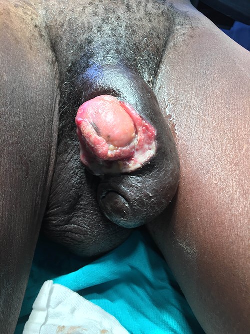
Upon arrival to MCH, the patient was hemodynamically stable with no systemic signs of infection. On physical examination, he was noted to have a 3 cm by 3 cm fungating mass on the right aspect of his penile shaft (Fig. 1). The patient also had firm, bilateral inguinal lymphadenopathy concerning for malignancy. Both the mass and inguinal areas were moderately tender to palpation. Our preliminary diagnosis at this time was penile cancer versus genital infection. We cleaned the mass and applied a sterile gauze dressing and continued with daily dressing changes. A complete blood count was ordered which revealed a microcytic anemia (hemoglobin 10.1 g/dl) and a normal white blood cell count (5400 cells/ul) without left-shift. He had no other systemic signs of infection, however due to inability to rule-out an infectious etiology, the patient was started on IV ceftriaxone.

Fungating penile mass emerging from mid-penile shaft with urinary leakage. Bilateral inguinal lymphadenopathy was also present on physical examination.
Surgical course
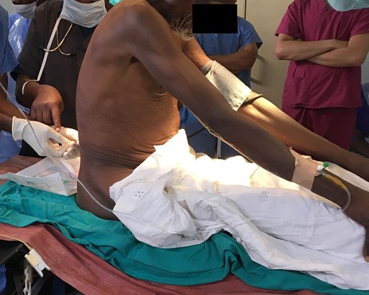
The patient was taken to major theater later that day for exploration and penile amputation. Spinal anesthesia was induced given that general anesthetic agents were running low and reserved for emergency cases (Fig. 2). After the patient’s penis and bilateral groins were prepped and draped, we then turned our attention to the penile mass. A circumferential incision was made through the skin at the base of the penis, away from the lesion itself. A skin flap was then raised circumferentially using electrocautery and blunt dissection. The penis was degloved, then a tourniquet was placed to ensure hemostasis. A penile amputation was performed and the specimen was sent for permanent pathology along with inguinal lymph node. The exposed corpora cavernosa were closed with a running 0-Vicryl suture. The tourniquet was then removed and hemostasis was ensured along the amputated surfaces.

Eighty two-year-old man undergoing spinal anesthesia prior to surgical resection of penile mass.
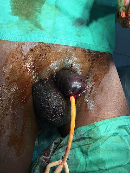
Next, we moved on to the reconstruction portion of the operation. Scrotal flaps were raised bilaterally by making a vertical incision into the median raphe. A left hydrocele was encountered which was opened, drained, and evaginated using absorbable sutures. The scrotal flaps were then used to perform a complex reconstruction by closing the flaps over the remnant urethra. The urethra was spatulated sharply along the three- and nine-o-clock aspects, then the spatulated opening was sewn to the scrotal flaps using absorbable sutures. A 20-French Foley catheter was gently inserted into the reconstructed urethra avoiding any undue tension. The remainder of the skin flap was closed in multiple layers, and the procedure was completed (Fig. 3).

Image of post-penectomy reconstruction with testicular flaps and Foley catheter drainage. Some wound separation is noted at the reconstruction margins.
Postoperative course
Postoperatively, the patient was taken to the post-anesthesia care unit for recovery, then transferred to the male surgical ward. He was continued on IV ceftriaxone to complete a seven-day course, which is standard protocol given the poor sterile conditions at MCH. Pain control was achieved with diclofenac and paracetamol. Surgical dressing was removed on the third postoperative date. We noted that the skin flaps had approximately 5% necrosis on the inferior aspect of the reconstructed penis, but otherwise appeared healthy without separated tissue. The patient was discharged on the seventh postoperative date after his family members were taught dressing change techniques. The patient’s Foley catheter was kept in place at discharge to allow for urethral healing without stricture formation, with planned removal at follow-up. Pathology report was not available at time of discharge.
DISCUSSION
We present the case of an 82-year-old man with a three-week history of a fungating penile mass with urinary leakage. Penile cancer presenting as a fungating mass with urinary leakage and infection is a rare but urgent condition that requires surgical intervention. The differential diagnosis of penile masses can be subdivided into three broad categories. These include inflammatory skin lesions, infectious lesions (e.g. genital herpes), and neoplastic lesions. In the present case, the mass had clearly invaded the penile urethra as evidenced by urinary leakage from the mass itself. This suggests an invasive neoplastic process, most likely a squamous cell carcinoma. The patient reported a three-week history of penile swelling and pain, but this process had likely been going on for a much longer period.
In a third-world setting where chemotherapy and radiation therapy are not available, management of such lesions is surgical. We present one surgical option for resection and reconstruction of an invasive penile mass. One should note that this does not represent a curative oncologic resection, given that the patient’s inguinal lymphadenopathy reflects a disseminated disease process. Given the patient’s age and available clinical resources, however, palliative resection is the patient’s best healthcare option available. Unfortunately, we do not have the pathology results available to us, nor has the patient followed up in our surgical clinic since discharge to evaluate wound healing status and recurrence.
In summary, we highlight the optimal management of a fungating penile mass in a third-world country with low healthcare resources. Neither chemotherapy nor radiation therapy are reliably available in this setting, so options are limited to surgical resection for palliation.
AUTHOR CONTRIBUTIONS
YK, MCM, TCL, and REE made substantial contributions to conception and design of the article, initial drafting, and critical revisions. All authors approve of the final version and agree to be accountable for the article.
CONFLICTS OF INTEREST
All authors have no conflicts of interest to disclose.


