-
PDF
- Split View
-
Views
-
Cite
Cite
Andrew Sweeny, Nathan A Smith, Jennifer A Serfin, Hemorrhagic cholecystitis causing hemobilia and common bile duct obstruction, Journal of Surgical Case Reports, Volume 2019, Issue 3, March 2019, rjz081, https://doi.org/10.1093/jscr/rjz081
Close - Share Icon Share
Abstract
Biliary obstruction is a common problem associated with gallbladder pathology. However, hemorrhagic cholecystitis with hemobilia as the cause is quite rare. We present a case of hemorrhagic cholecystitis in the setting of systemic anticoagulation causing common bile duct obstruction which required endoscopic retrograde cholangiopancreatography (ERCP) for ductal clearance followed by laparoscopic cholecystectomy. The triad of right upper quadrant abdominal pain, jaundice and gastrointestinal bleed should prompt consideration of hemobilia in the setting of biliary obstruction.
INTRODUCTION
Hemorrhagic cholecystitis is a rare condition with a wide spectrum of potential presentations. Hemobilia has a well-described triad of abdominal pain, jaundice, and gastrointestinal bleeding which is present in approximately 22% of cases [1]. Since anticoagulant agents are used by a wide variety of patients, hemorrhagic cholecystitis should be considered when unusual presentations of cholecystitis are encountered. We present a case of hemorrhagic cholecystitis causing biliary obstruction as well as a brief review of published case reports.
CASE PRESENTATION
A 78-year-old male with past medical history of atrial fibrillation (on Warfarin), Type 2 diabetes mellitus, hypertension, and coronary artery disease presented to the Emergency Department with a chief complaint of epigastric abdominal pain radiating to the central abdomen which was worsened with food intake. Associated symptoms included nausea, emesis, fever, and chills. On physical exam, he was noted to have epigastric tenderness, absent Murphy’s sign, scleral icterus and an irregularly irregular heart rhythm. He did not demonstrate symptoms of gastrointestinal bleeding at the time of presentation.
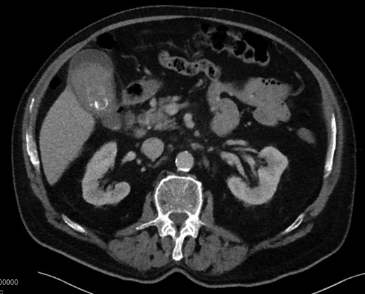
Laboratory results were as follows: white blood cell count 10.5, hemoglobin 13.9, platelet count 164, Total bilirubin 3.8, AST 133, ALT 200, alkaline phosphatase 339, lipase 33, protime 36.3, INR 4.2. Abdominal ultrasound was obtained to evaluate for potential gallbladder/biliary pathology given his presentation. The ultrasound demonstrated gallstones with findings concerning for chronic cholecystitis with a common bile duct measurement of 8 mm. To further evaluate the cause for the patient’s abdominal pain, a computed tomography (CT) scan was performed of the abdomen and pelvis. CT findings demonstrated an abnormal gallbladder with stones and dense intraluminal debris measuring 53 Hounsfield units. Similar debris was seen in the common bile duct with a measurement of 17 Hounsfield units. Active contrast extravasation into gallbladder lumen was not seen on the CT, but Hounsfield units were consistent with intraluminal blood. The common bile duct was measured at 8 mm in diameter without intrahepatic ductal dilatation (Fig. 1).

CT with dense heterogeneous gallbladder contents and cholelithiasis.
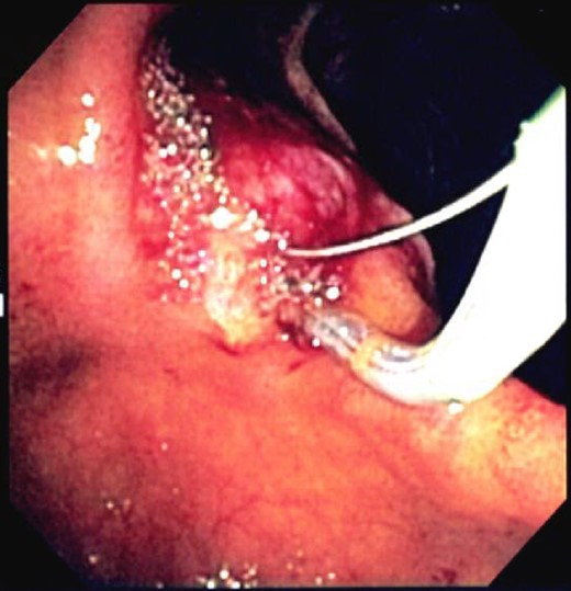
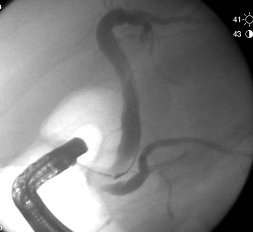
The patient was admitted for management of obstructive jaundice with possible cholecystitis. On hospital day 1 his bilirubin increased to 5.1, he was given Vitamin K and fresh frozen plasma to correct his coagulopathy and he was taken for endoscopic retrograde cholangiopancreatography (ERCP). During ERCP, the common bile duct (CBD) was cannulated and swept revealing a moderate amount of maroon clot (Fig. 2). No other debris or stones were noted to be within the CBD on final fluoroscopic image (Fig. 3).

Endoscopic image of ERCP with hemorrhagic contents being removed.

On hospital day 2, laboratory values improved overall with bilirubin of 2.1, AST 57, ALT 130 and Alkaline phosphatase of 254. The patient was then taken to the OR for laparoscopic cholecystectomy. Intraoperative findings included: dense omental adhesions, thickened gallbladder wall, extensive pericholecystic edema, cholelithiasis, and a large clot within the gallbladder lumen. Postoperatively, patient’s recovery was uneventful with down trending direct bilirubin, resolution of pain, and successful restarting of his Warfarin. He made a full recovery by his post-operative follow-up visit.
DISCUSSION
Hemorrhagic cholecystitis is a rare cause of hemobilia and an even more rare etiology of biliary obstruction. Non-iatrogenic causes of hemobilia include blunt trauma, anticoagulation, renal failure, cirrhosis and cancer [2]. In cases of hemorrhagic cholecystitis, bleeding into the gallbladder lumen is caused by transmural inflammation of the gallbladder wall leading to infarction, erosion, and bleeding [3]. Hemobilia has a described triad of presenting symptoms, including right upper quadrant pain, jaundice and GI bleeding [4].
Few case reports have been published describing the presentations and treatment modalities of this disease process. After an extensive literature search, we were able to identify two similar cases to ours with some variations. One case report published by Dong Keun Seok described a patient presenting with pain, jaundice and symptoms of GI bleed. Active contrast extravasation into the gallbladder was noted on CT without evidence of stones. Intra-operative findings were consistent with arterial bleeding into the gallbladder lumen [5]. Another case report described a similar presentation without signs of active hemorrhage on CT. Cholecystectomy was performed which resolved the hemobilia as well as the obstructive process [4].
The work-up for hemorrhagic cholecystitis, as with any suspected gallbladder pathology, should start with history, physical, labs and imaging. Imaging findings may be able to distinguish acute calculous cholecystitis from hemorrhagic cholecystitis. Ultrasound findings of hemorrhagic cholecystitis can show gallbladder wall thickening, intraluminal membranes, and non-shadowing, non-mobile intraluminal echogenic material [6]. CT findings may demonstrate contrast extravasation, high attenuation within the gallbladder lumen and fluid-fluid layering [7].
The common themes noted in the case reports as well as with our patient are to remove the gallbladder and to relieve the biliary obstruction if present, through use of ERCP. In our patient, anticoagulant therapy played a large role in his bleeding. Unfortunately, there are no large studies or epidemiologic data to demonstrate the true incidence of hemobilia caused by anticoagulant therapy or by hemorrhagic cholecystitis; more investigation is required to answer this question.
CONFLICT OF INTEREST STATEMENT
None declared.


