


Abstract
Penetrating abdominal trauma is an uncommon cause of presentation to emergency departments in Australia and is frequently associated with the clinical need for emergent operative intervention. Advances in imaging modalities, improved laparoscopic techniques and structured approaches to resuscitation in trauma have now allowed potential minimally invasive management of such injuries, avoiding laparotomy and therefore defining peritoneal breach; the major determinant of intra-abdominal organ injury in this setting is critical. We present the case of a self-inflicted stab injury to the suprapubic region in an otherwise healthy man and describe the combination of imaging and operative modalities used to define peritoneal breach in this case which successfully reduced the patient’s morbidity by avoiding non-therapeutic laparotomy.
INTRODUCTION
Defining peritoneal breach in patients with penetrating abdominal trauma can be difficult as reliance on clinical examination and imaging alone may not be sufficient in all cases, and progressing directly to laparotomy is associated with a high risk of non-therapeutic intervention. We present the case of a self-inflicted stab wound to the suprapubic region, using a combination of imaging and operative techniques to define the breach and avoid non-therapeutic laparotomy.
CASE REPORT
A 67-year-old male presented to the emergency department with an alleged self-inflicted stab wound to his suprapubic region, having been found alone in a public toilet by a passer-by surrounded by an unknown volume of blood. Past medical history included a coronary artery bypass graft on aspirin, depression and benign prostatic hypertrophy. He described stabbing himself with a knife as a method of alleviating protracted rectal pain.
He was managed according to Emergency Management of Severe Trauma (EMST) guidelines, with initial treatment based on resuscitation from expected blood loss. His haemodynamic measurements were within the normal range throughout his transfer and resuscitation. Physical examination revealed a patient in acute pain and a knife, directed in an infero-posterior direction in the midline of the abdomen, 1–2 cm inferior to the intertubercular plane [1], surrounded by a clot and an expanding haematoma of the mons pubis (Fig. 1). Despite being generally blood-soaked, no blood was expressible from the urethral meatus. No other penetrating injuries were identified. Biochemistry revealed lactate of 4.7 mmol/l, normal pH and creatinine, a white cell count 9.9 × 10^9/l and haemoglobin 140 g/l. An extended focussed ultrasound in trauma (eFAST) was performed, which showed no abdominal free fluid. The knife was left in situ and stabilised with a bolster of combine dressings and tape.
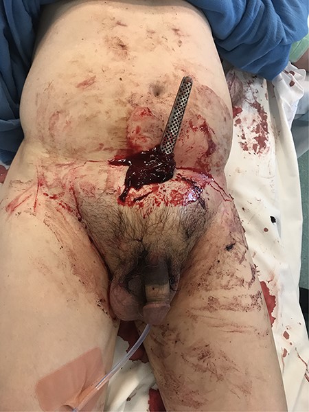
Photograph of the stab wound displaying the infero-posteriorly directed knife and surrounding haematoma of the mons pubis. Bruising can be seen extending to the penis.
After discussion with urology, a 14Fr indwelling catheter was inserted, which drained yellow urine. A computed tomography (CT) mesenteric angiogram demonstrated penetrating injury to the suprapubic region with haematoma and two foci of arterial extravasation with radiological evidence of peritoneal breach, and the metallic foreign body in situ (Figs 2 and 3). Importantly, the tip of the knife appeared to end at the base of the penis at the expected confluence of the dorsal vein.
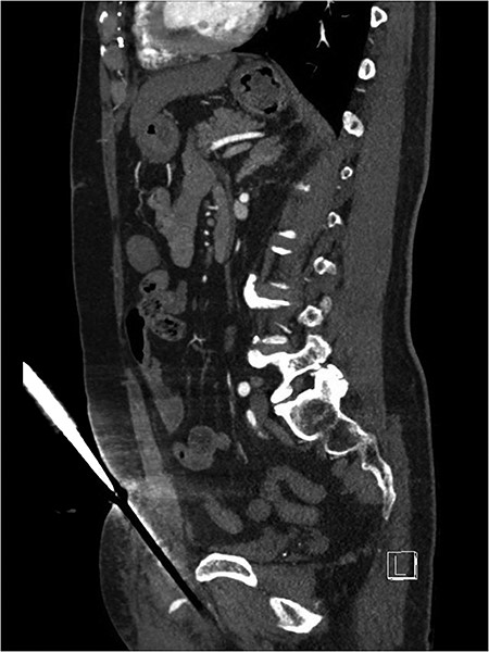
Suprapubic stab wound: sagittal section: CT mesenteric angiogram showing knife directed in infero-posterior direction abutting but not traversing the peritoneum. Active arterial contrast extravasation can be seen. The tip of the knife abuts the root of the penis.
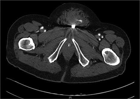
Suprapubic stab wound: coronal section: CT mesenteric angiogram showing mons pubis haematoma with active arterial extravasation.
He underwent an urgent exploratory laparoscopy which showed no evidence of peritoneal defect and no intra-abdominal blood. The knife was removed and placed in a sealed bag at police request. The wound was extended transversely and explored, defining a cavity extending to the pubic symphysis with active bleeding from rectus muscle controlled with diathermy. The cavity was debrided and irrigated, and FLOSEAL was applied. The wound was closed primarily. He had a postoperative haemoglobin of 124 g/l, and an uncomplicated admission, and he was referred to Psychiatry for ongoing management.
DISCUSSION
Penetrating abdominal injuries are an uncommon cause of trauma presentation in New South Wales, representing 1.1% of all-cause trauma admissions with self-harm representing 3.4% of all mechanisms, with a high associated case fatality rate for those severely injured (8.2% of trauma-related deaths) [2]. The management of penetrating abdominal injuries with radiological evidence of intra-abdominal free fluid and haemodynamic instability has been well defined in the EMST guidelines; however, there is a subset of false-negative eFAST scans, who may also have a negative CT but still have an intra-abdominal injury or peritoneal breach. A recent retrospective review of haemodynamically stable patients with penetrating abdominal trauma found that 25% had intra-operative findings despite a negative CT scan [3]. CT tractography may define peritoneal breach better than conventional CT; however, its high rate of false negatives limits its use [4]. Evidence in this cohort is therefore lacking, and local protocols exist, which direct towards non-operative management, wound exploration alone, laparoscopy, laparotomy or a combination of these methods.
In our trauma referral hospital, in patients with negative imaging and the absence of haemodynamic instability, our preferred method is a diagnostic laparoscopy to first define the presence of intra-abdominal injury and peritoneal breach. This reflects recent literature suggesting that diagnostic laparoscopy may be considered a tool to evaluate for peritoneal injury and reducing non-therapeutic laparotomies in haemodynamically stable patients [5–8]. Rates of non-therapeutic laparotomy as high as 25% have been described [9]. Progression to laparotomy would then be both injury severity and surgeon dependent, as the operation may be completed successfully via laparoscopy. A recent Australian review highlighted that peritoneal breach alone as an indicator for laparotomy is associated with a moderate incidence of non-therapeutic laparotomy [10]. Our approach intends to reduce the rate of non-therapeutic laparotomy, which increases patient morbidity in both the short- and long-term through postoperative pain, ileus, wound infection, bowel injury, small bowel obstruction and hernia risk.
CONCLUSION
We have described a case of self-inflicted penetrating lower abdominal injury that despite being extra-peritoneal, has highlighted the importance of a protocolled approach to trauma patients to objectively define their injury and to tailor their operative intervention thereby reducing their iatrogenic morbidity.
Conflict of Interest
There are no conflicts of interest to declare.
All authors are in agreement with the content of the manuscript.
Consent for the report has been granted by the patient.
The manuscript has not been published previously and is not under consideration elsewhere.
