


Abstract
Internal hernia of the small bowel is a rare finding especially in previously non-operated abdomen. Such a hernia occurring due to involvement of appendices epiploicae is an even rare instance with less than five reported cases in the literature. We encountered a 75-year-old male who had internal herniation of small bowel through an aperture created by adhesion between two appendices epiploicae in a previously virgin abdomen. Laparotomy and division of adhesion was performed to manage him successfully. Even in a virgin abdomen, a high index of suspicion along with early intervention is the key to reduction in mortality and morbidity in cases intestinal obstruction.
INTRODUCTION
Intestinal obstruction occurring due to internal herniation of bowel is very rare. Internal herniation occurring due to appendices epiploicae is even more uncommon with less than five reported cases in literature [1, 2]. Epiploic appendages or appendices epiploicae are peritoneum-lined protrusions of subserosal fat that arise from the surface of the colon. Inflammation of these appendices can cause intestinal obstruction by forming a band due to fusion [3].
We present here a rare case of a 75-year-old man who came to us with intestinal obstruction in a previously non-operated abdomen. Radiological investigation indicated obstruction although no cause could be identified. He was explored and found to have an internal herniation from an aperture formed by two adhered appendices epiploicae. Division of the adhesion was successful to relieve the obstruction and no resection was required. Early intervention even in previously virgin abdomens carries great importance for a successful outcome in intestinal obstruction.
CASE PRESENTATION
A 75-year-old male patient presented with inability to pass stools and flatus with recurrent vomiting and progressive abdominal distension for 3 days. He was mildly dehydrated and anxious. His vitals showed a tachycardia of 100 /m. Blood pressure and temperature was normal on presentation. Physical examination showed abdominal distension without any visible peristalsis and organomegaly. He was diagnosed as a case of intestinal obstruction. His blood parameters were within range. Radiological investigation including a CECT of the abdomen revealed small bowel obstruction with a clear cut off. However, the cause of obstruction could not be identified.
He underwent exploratory laparotomy wherein intestinal obstruction involving the proximal ileum was found. This obstruction was due to herniation of a 20 cm loop of ileum through an aperture created by adhesion between two appendices epiploicae of the sigmoid colon.
Release of the adhesion was done followed by epiploicae appendectomy of the involved epiploicae. Bowel showed only early signs of vascular compromise and no resection was required. Postoperative phase was uneventful and patient was discharged on the third postoperative day.
DISCUSSION
Appendices epiploicae are peritoneum-lined protrusions of the subserosal fat that arise from the surface of the colon. They do not have a known function. These typically measure 1.5 × 3.5 cm2 but have been reported to measure up to 15 cm in length. There are between 50 and 100 of them in the large bowel and extend from the caecum (where they may be absent) to the rectosigmoid junction. They are distributed longitudinally in two rows on the medial (along the taenialibera) and the posterolateral (along the taeniaomentalis) aspects of the large bowel. There is only one row of appendices epiploicae along the transverse colon and there are none along the rectum [4]. Pathological conditions occurring because of these are very rare. An internal herniation involving these appendices can occur because of inflammation causing adhesions between appendices and adjoining structures or between two appendices themselves. Such an inflammation may even occur in a virgin abdomen (Fig. 1).
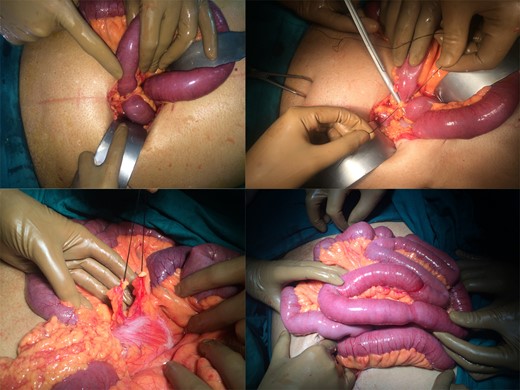
Operative pictures.
Internal hernias, as such are very rare and intestinal obstruction occurring due to internal hernia account for 0.2–0.9% only [5, 6]. No radiological investigation is helpful in diagnosis an internal hernia and a high index of suspicion with timely intervention is lifesaving. Radiology in cases of intestinal obstruction can only help to prove the diagnosis and indicate the level of obstruction; however, the cause of obstruction is rarely identified on radiology. In a case of herniation in a mesenterial aperture the treatment consist of closing the opening after reducing the hernia. In the case described above release of adhesion with epiploicae appendectomy will suffice.
To the best of our knowledge, an internal herniation because of an adhesion between the tips of two appendices epiloicae has never been reported before. We found less than five cases of internal herniation caused by adhesions between appendicices epiploicae and surrounding omentum [1, 2]. We presume that in this patient who was previously not operated, mild inflammation in the appendices must have lead to adhesions and herniation.
CONFLICT OF INTEREST STATEMENT
None declared.
