-
PDF
- Split View
-
Views
-
Cite
Cite
Brenton Robinson, Rapid resolution of severe subcutaneous emphysema with simple percutaneous angiocatheter decompression, Journal of Surgical Case Reports, Volume 2018, Issue 7, July 2018, rjy173, https://doi.org/10.1093/jscr/rjy173
Close - Share Icon Share
Abstract
Subcutaneous emphysema (SE) is often seen as a sequela of chest tube placement, cardiothoracic surgery, trauma, pneumothorax, infection or malignancy. In most cases SE is self-limited and requires no intervention. Rarely, air can rapidly dissect into subcutaneous tissue planes leading to respiratory distress, patient discomfort and airway compromise. This is a case of a 75-year-old woman that developed massive SE and impending respiratory failure with rapid progression of air into her subcutaneous tissue. In an effort to rapidly stabilize the patient we placed multiple percutaneous angiocatheters into the subfascial space with complete resolution in <24 h. This technique was an excellent temporizing measure and found to be superior to previously described techniques involving large open ‘blow hole’ incisions or large bore drains. Placement of angiocatheter needles for the decompression of subcutaneous air is a well-tolerated, readily accessibility, low cost and simple procedure for the treatment of SE.
INTRODUCTION
Subcutaneous emphysema (SE) is a relatively frequent entity seen in the intensive care unit that under most circumstances spontaneously resolves. However, depending upon the extent and acuity of SE there are situations in which rapid progression with subcutaneous air tracking into multiple tissue planes can cause severe patient discomfort, airway compromise, cardiac tamponade and tension pneumomediastium [1]. Massive accumulation of air in the deeper tissue spaces at the level of the thoracic outlet has the capability of compressing the trachea and great vessels thereby severely compromising the airway, venous return, and blood flow to the head and neck [2]. Etiology can be the end result of a number of processes including; blunt or penetrating trauma, pneumothorax, infection, malignancy, or as a complication of surgical procedures and in rare cases spontaneous SE may be seen [3]. The most common clinical sign of SE is the characteristic diffuse tissue distention classically presenting with marked palpable cutaneous tension and crepitation on palpation. As SE progresses subsequent ventilator requirements and specifically ventilator pressures required to maintain adequate oxygenation can leave practitioners stuck in a clinical conundrum of progressively increasing inspiratory pressures at the expense of worsening SE. Various approaches have been described in the literature to alleviate the pressure including the use of open blowhole incisions, negative pressure wound therapy, drains or cervical mediastinotomy [1, 4–6].
CASE PRESENTATION
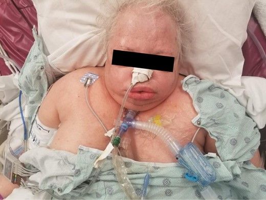
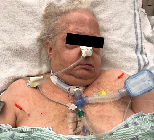
A 75-year-old female was initially admitted to the ICU after planned esophagogastrectomy for esophageal cancer with laparoscopic mobilization of stomach and duodenum by a left posterior lateral thoracotomy and subsequent gastric pull up and reconstruction. Patients esophageal cancer was a partially obstructing and circumferential T4N1 (stage IIIA) moderately differentiated adenocarcinoma at the gastroesophageal junction. Intraoperatively, the patient’s visceral pleura was violated necessitating the placement of two 24 Fr chest tubes. Upon subsequent chest X-ray (CXR) imaging 5 days after removal of the initial chest tubes it was noted that there was recurrence of a small apical pneumothorax and a percutaneous chest tube was subsequently placed. Post procedure imaging revealed the percutaneous tube was placed directly into the parenchyma of the right lung creating a large air leak. Subsequently an open chest tube was placed at bedside and 40 ml of blood was evacuated. Shortly after the passage of the new chest tube and the patient developed increasing ventilator requirements the patient began developing clinical signs of SE. Physical exam revealed widespread upper trunk tissue distention and crepitance to palpation. Within hours widespread tissue distention spread cephalad to involve the upper chest, neck and face. The patient progressively developed massive SE and increasing peak pressures (Fig. 1). The following day the patient developed complete inability to open her eyes and her peak airway pressures became elevated to 70 cm H2O. The patient had a massive and persistent air leak and negative pressure was increased in the chest tube drainage system to −40 mmHg without any improvement in respiratory dynamics. In an effort to rapidly decompress the rapidly evolving SE we decide to place three 14 g 2 inch I.V. catheters (Acuvance® Plus Safety I.V. Catheter—Smiths Medical ASD, Inc. Southington, CT) into the tissue. The first needle was placed at the area of the greatest air accumulation in the right upper lateral chest wall (Fig. 2). The trajectory of the needle was into the deep plane of the chest wall. Upon successful placement of the needle there was audible release of air immediately decompressed. Subcutaneous air subsequently continued to decompress and was noted to be in synchrony with the respiratory cycle. Lateral to the initial needle another 14 g angiocatheter was placed in an oblique and transverse direction in an effort to provide a drainage site to a more superficial tissue plane (Fig. 3). Lastly a third 14 g needle was placed on the contralateral left upper lateral chest wall directed in a deep oblique direction (Fig. 2). During the placement of all three needles there was an audible rush of air that was evacuated and continued to decompress once in final position. After successful placement, the inner needle was removed and the overlying plastic angiocatheter was left in place. Within the first three hours there was a noticeable change in both a decrease in the tissue distention and improvement in respiratory dynamics. The patient continued to progressively improve over the initial 24 h with near complete resolution of SE and normalization of ventilator dynamics, most notably in normalization of peak airway pressures indication alleviation of airway compression. The angiocatheters were subsequently left in place for 24 h and removed without difficulty. Measuring from the level of the skin to the proximal hub of the catheter proved to be a simple means to quantify the degree of decompression and in this patient, we were able to achieve 3 cm of circumferential decompression (Fig. 4). At 24 h, there was nearly complete resolution of SE (Fig. 5).
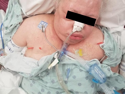
Angiocatheter placement into the right upper lateral chest wall and left upper chest wall.
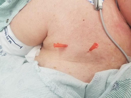
Angiocatheter placement into the deep and oblique fascial layers.
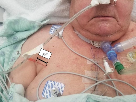
24 hours after placement of angiocatheter demonstrating 3 cm of circumferential decompression from the level of the skin to the proximal hub of the catheter.
DISCUSSION
In review of the literature we found a small number of case reports highlighting techniques to aid in alleviating the pressure in SE; blowhole incisions, negative pressure wound therapy, drains or cervical mediastinotomy. While all of the techniques are effective in decompressing and relieving the subfascial pressure we believe that our technique is advantageous because it is minimally invasive, simple and effective. This technique requires no special drains or equipment and does not have large open infraclavicular incisions which have been associated with bleeding, insufficient depth and poor cosmesis [3].
In practice, this technique has proven to be an excellent temporizing measure to aid in the complete resolution of the underlying etiology of SE, or effectively as a bridge to a more definitive procedure like a video assisted thoracoscopic wedge resection of the affected segment of lung. The placement of the angiocatheter needles is a well-tolerated procedure because the massive distention of the skin renders the skin relatively insensate. Lastly, the easy accessibility, low cost, simple and quick application and high efficacy are all particularly advantageous components in the use of percutaneous angiocatheters for the treatment of severe SE.
CONFLICT OF INTEREST STATEMENT
None declared.


