-
PDF
- Split View
-
Views
-
Cite
Cite
Rahil Dharia, Vinu Perinjelil, Rohit Nallani, Fadi Al Daoud, Gul Sachwani-Daswani, Leo Mercer, Kristoffer Wong, Superficial femoral artery transection following penetrating trauma, Journal of Surgical Case Reports, Volume 2018, Issue 6, June 2018, rjy137, https://doi.org/10.1093/jscr/rjy137
Close - Share Icon Share
Abstract
We describe a patient who sustained a penetrating injury to the posterior right lower extremity just above the popliteal region with transection of the superficial femoral artery (SFA) despite minimal evidence of active bleeding. An on-table angiogram identified flow in the SFA followed by the popliteal artery and into the trifurcation of the right lower extremity. Eventually, a second operation revealed transection followed by end-to-end anastomosis of SFA and stabilization of the patient. The findings of this case highlight the need for a high index of suspicion and persistent clinical investigation to identify vascular injuries in the absence of hard signs of vascular trauma.
INTRODUCTION
Transection of the superficial femoral artery (SFA) following blunt or penetrating trauma is rare, in contrast to several reported cases of SFA injury from fractures and iatrogenic etiologies. Transection of SFA can lead to significant hemodynamic instability and long-term sequelae if not quickly identified and adequately addressed. Penetrating injuries comprise 90% with gunshot wounds representing most common mechanism. We present a case of SFA transection following a stab wound with an atypical bleeding pattern that was managed with primary repair and emphasize different management approaches in the setting of vascular trauma.
CASE REPORT
A 28-year-old female presented after sustaining multiple stab wounds to bilateral arms, and posterior right thigh. The patient was hypotensive with a blood pressure of 67/42 and tachycardic with a pulse of 140 in the emergency department with easily palpable carotid and radial pulses but intermittent dorsalis pedis pulses. She had a large posterior thigh wound with active bleeding. A tourniquet was placed at the thigh followed by initiation of massive transfusion protocol and she was promptly taken to the operating room (OR) for exploration of the right thigh wound.
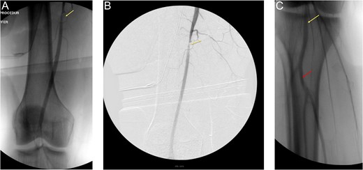
In OR, a horizontal groin incision was performed with dissection carried down to the right femoral artery and vein. After proximal control was achieved, intra-operative Doppler ultrasound of the popliteal, posterior tibial and dorsalis pedis vessels yielded triphasic signals. An on-table angiogram through the right common femoral artery depicted flow through the SFA and profunda femoral arteries with no active extravasation (Fig. 1A and B).

(A) and (B) are on-table angiograms depicting flow through SFA, without any contrast extravasation. (C)— On-table angiogram depicting flow from popliteal artery to trifurcation, without any contrast extravasation.
Although there was filling defect noted on Fig. 1B, contrast was shown flowing to the popliteal artery and thereby into the trifurcation of the right lower extremity (Fig. 1C).
It was noted that the posterior thigh wound was not actively bleeding and was packed with hemostatic agent.
As the patient was being prepared for extubation, right posterior thigh dressings were saturated and bleeding was identified from the wound. The patient became hypotensive. This wound was packed and second exploration was performed with patient in prone position to provide better exposure. When the packing was removed, active bleeding was seen and vascular clamps were applied to proximal and distal segments of a branch of the SFA. Doppler showed audible signals in the popliteal as well as posterior tibial artery. After the bleeding artery was suture ligated as well as clipped, hemostasis was obtained. A post-operative CT angiogram (CTA) of the right lower extremity was performed revealing occlusion of the SFA proximal to the adductor canal. Upon re-exploration and clearing a substantial amount of hematoma occluding the SFA, patient was found to have complete SFA transection with each end retracted. The injury was surgically managed with end-to-end anastomosis. Post-operatively, patient had Doppler dorsalis pedis and posterior tibial signals. Follow-up in clinic demonstrated palpable pulses in bilateral lower extremities.
DISCUSSION
Femoral vessel injuries account for approximately 70% of all extremity vascular injuries, most incurring from penetrating mechanisms [1]. Arterial injuries can be categorized as partial transections, complete transections, contusions with thrombosis, intimal disruption and arterio-venous fistula. Complete transections can lead to vasospasms on each end with minimal bleeding minimizing the suspicion for major vascular injury [2]. There is an increased potential for missed vascular injuries in the absence of hard signs of vascular trauma after penetrating mechanism. In the presence of ‘hard’ signs (pulse deficit, distal ischemia, expanding hematoma, arterial bleeding), angiogram and surgical management of injury is appropriate. Judicious assessment of these ‘hard’ signs can be critical in limb salvage. In the presence of ‘soft’ signs (stable hematoma, wound proximity to major vessel, unexplained hypotension), management can be difficult and occult vascular injury can be missed. Complications of missed vascular injuries can lead to lifelong functional disability or even loss of limb [3].
Measurement with Ankle–Brachial Index (ABI) is a non-invasive and reliable screening method to rule out vascular injuries in hemodynamically stable patients. When ABI is <0.9, further work up can be done with CTA or conventional angiography. Angiography is the modality of choice in assessing the extent of damage and aiding in its’ surgical management. Pre-operative CTA if performed, can show partial or total occlusion, active extravasation, pseudo-aneurysm, intimal flaps and arterio-venous fistulas. When CTA is inconclusive, a conventional angiography can be performed [4]. Even in the absence of overt signs of arterial injuries, arteriogram should be performed to evaluate the potential for vascular injury, when intra-operative Doppler assessment is negative or inconclusive. Multiple angiographic angled-views of the site of injury should be undertaken to not miss an occult partial transection or dissection flap. Patients with early missed vascular injuries present with recurrent hemorrhage as in this case and late missed injuries can be complicated with pseudo aneurysms, arterio-venous fistulas, arterial occlusion, etc [5]. Injuries to SFA can be managed with primary repair including end-to-end anastomosis, interposition saphenous vein/PTFE graft, vein patch or femoro-femoral bypass with reversed saphenous vein or PTFE conduit. All arterial injuries are managed with debridement, thrombus clearance by Fogarty catheter, and definitive repair. Goals during revascularization are tension free anastomosis and adequate soft-tissue coverage with liberal use of fasciotomies after repair. Selection of an autologous saphenous vein from the contralateral side is suitable and when the selected vein is less than 6 mm, a synthetic graft can be used instead [6]. In cases with co-existing fractures, reports of successful management of arterial injuries by stenting allow for reperfusion distally and definitive management of co-existing injuries. Vascular injuries have increased propensity to be missed if there are no co-existing injuries and missed diagnosis for greater than 6 h can lead to limb compromise. The use of temporary shunts can play a vital adjunct in damage control prior to definitive repair of the vessel. In cases where temporary shunted injuries were then surgically repaired with interposition grafts, there was a reported 92% viability in those extremities [7].
Penetrating injury to femoral artery can have significant morbidity and mortality but a high index of suspicion without the presence of overt signs of bleeding should invoke further investigation in prevention of associated complications.
CONFLICT OF INTEREST STATEMENT
None declared.


