-
PDF
- Split View
-
Views
-
Cite
Cite
Dionysia Vasdeki, Effrosyni Bompou, Alexandros Diamantis, Athanasios Anagnostou, Konstantinos Tepetes, Matthaios Efthimiou, Haemangiopericytoma of the greater omentum: a rare tumour requiring long-term follow-up, Journal of Surgical Case Reports, Volume 2018, Issue 5, May 2018, rjy087, https://doi.org/10.1093/jscr/rjy087
Close - Share Icon Share
Abstract
Haemangiopericytomas (HPC) are highly vascularized tumours located in any part of the body where capillaries can be found. Since 2002, they have been re-classified under the umbrella ‘extrapleural Solitary Fibrous Tumour (SFT)’ and the term HPC is nowadays used to describe a growth pattern rather than a clinical entity. Their biological behaviour varies and they require a long-term follow-up since they may recur or metastasise several years after successful treatment. We present the case of a gentleman with HPC of the greater omentum initially appeared in 1998. HPC rarely develops in the greater omentum and only 20 cases have been described in the literature till today. Despite complete excision the mass re-appeared in 2011 and 2017, 13 and 19 years after initial treatment. Surgical management included en bloc excision of three lesions along with greater omentum. No further treatment was required.
INTRODUCTION
Haemangiopericytomas (HPC) are highly vascularized tumours located in any part of the body where capillaries can be found [1]. They were first described in 1942 and since 2002 they have been grouped into the umbrella ‘extrapleural solitary fibrous tumour (SFT)’ [2]. Their biological behaviour varies and long-term follow-up is required, even after successful management [3]. We present the case of a gentleman with HPC of the greater omentum initially appeared in 1998 and, despite successful management, it recurred in 2011 and 2017.
CASE REPORT
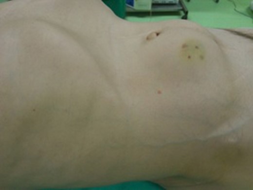
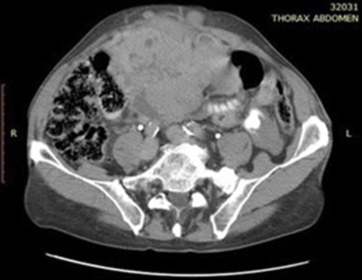
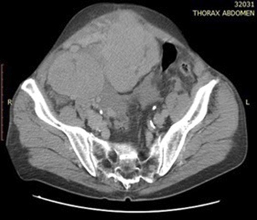
A 72-year-old gentleman presented to our unit with a recurrent mass of the anterior abdominal wall. This mass appeared gradually over several months causing a dull pain. No other symptoms were described. Clinical examination revealed a 11 × 10 cm soft and well defined, palpable mass on the right side of the abdominal wall (Fig. 1). Computer tomography (CT) of the abdomen presented several space-occupying lesions in the peritoneal cavity, the right side of the lower pelvis and the rectovesical pouch, measuring 11 × 10.4 × 10.7 cm, 8 × 6.5 cm and 7.5 × 5.7 cm, respectively (Figs 2 and 3). Due to compression, the veins of the anterior abdominal wall presented dilated. Thorax CT scan revealed no tumour.

The patient presented with a 11 × 10 cm soft and well defined, palpable mass on the right side of the abdominal wall.


Abdominal CT scan showing two hypervascular lesions in the abdomen.
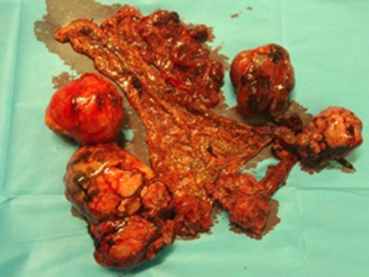
On exploratory laparotomy four large lesions were revealed arising from the greater omentum. No peritoneal or organ metastasis were seen. The lesions were excised en bloc with the greater omentum (Fig. 4). The masses were surrounded by blood vessels of large calibre, approximately 0.5 cm. Due to their vascular nature, there was a significant amount of blood loss intra-operatively and therefore the patient remained in ICU for 24 hours post-operatively. Subsequently he had an uneventful recovery and was discharged 7 days after his operation.

Intra-operatively three lesions derived from the greater omentum were excised along with the greater omentum.
The patient had undergone excision of similar lesions from the anterior abdominal wall in 1998 and 2011 (19 and 6 years before the present case, respectively). Both operations were performed in other institutions and only the biopsy reports were available to us. The first biopsy confirmed haemangiopericytoma, more likely malignant, due to display of pleomorphism, hypercellularity and infiltration of the capsule of the mass. In 2011, two lesions were excised, along with part of the greater omentum. Histological evaluation showed haemangiopericytoma. Immunohistochemical analysis showed positivity for vimentin, CD34 and CD99 and a mitotic count of 1 mitosis per 10 high-power fields (HPF). Biopsy of the present case also confirmed a haemangiopericytoma; necrosis was present, while mitoses were <4/10HPF.
DISCUSSION
Haemangiopericytoma has been considered a controversial highly vascularized tumour. It was first described by Stout and Murray in 1942 as a neoplasm derived from the pericytes of Zimmerman, modified smooth muscle cells that regulate the calibre of the capillary lumen [4, 5]. Since 2002 haemangiopericytomas have been reclassified as solitary fibrous tumour (SFT), since the term HPC gathers numerous non-related entities that share certain morphologic characteristics. The term ‘extra-pleural SFT’ describes extra-meningeal SFTs, haemangiopericytomas, lipomatous haemangiopericytomas and giant cell angiofibromas, as defined by the World Health Organization (WHO) [2, 6–8].
The term haemangiopericytoma is nowadays used to describe a growth pattern rather than a clinical entity [5]. Histologically, HPC are sinusoidal vascular tumours with staghorn-shaped blood vessels surrounded by spindle shaped cells [4]. Several tumour types share similar vascularity, resulting in difficulties in their diagnosis, management and prognosis. Immunochemistry has not met great success in the diagnosis of HPC, since cell markers present in normal pericytes, such as desmin and muscle actins, are not frequently found in HPC cells [4]. Moreover, they display several features of pericytic, fibroblastic and myofibroblastic differentiation, while they express CD34, CD99 and Bcl-2 antigens [5].
Soft tissue SFTs represent <2% of all soft tissue tumours; malignant HPCs represent <1% of all vascular tumours and around 5% of all sarcomatous tumours [2, 5, 6]. HPCs can more frequently be located at the lower extremities (34,9%), retroperitoneum and pelvic cavity (24,5%) [1, 9]. The greater omentum has been a rare site for its occurrence; only 20 cases of HPC of the greater omentum have been described [8]. The median age of diagnosis is 45 years. The distribution is equal in both sexes, while there is no evidence of increased familial incidence [1, 6, 9]. They can present varied biological behaviour from being a slow growing tumour to a mass with aggressive growth. They can metastasise to lungs, while recurrence and metastasis can develop several years after treatment. Therefore, a long-term follow-up is required even after radical excision of the mass [3].
The majority has a relatively indolent behaviour with presenting symptoms being vague for several months and not specific. Pain or abdominal fullness can be late symptoms associated with an enlarging mass or perineural invasion [2, 4, 7, 9].
The criteria for malignancy remain a topic of debate. Enzinger and Smith proposed large lesion size (>5 cm), high cellularity, high mitotic count (≥4/10 HPF), display of pleomorphism and the presence of tumour haemorrhage and necrosis as criteria indicating malignancy [1, 8, 9]. Pasquali et al.[2] presented that high mitotic rate, hypercellularity and pleomorphism have been associated with a higher risk of recurrence, approximately 50% at 5 years, while tumour size has been inversely associated with prognosis.
Treatment options of HPC cases should be discussed in a team with experience in sarcoma surgery. En-bloc surgical excision has been the mainstay of treatment, the aim being to achieve complete resection with negative margins. Radiation therapy can improve local control rates post-operatively, but cannot result in complete remission of HPC. It can be recommended for tumours displaying criteria for malignancy and as an alternative treatment for unresectable or recurrent HPC. There is little evidence to support the use of adjuvant chemotherapy in their management; partial or short-term remission of metastatic disease has been sporadically documented after chemotherapy [1, 2, 6, 9]. Tyrosine kinase inhibitors could provide long-lasting stable disease, although the experience is limited and more studies still need to be conducted [6]. The risk of perioperative bleeding has always to be considered. Their extreme vascularity can make some lesions unresectable due to high risk of significant blood loss. Pre-operative vascular embolization of the lesion has been an effective measure to reduce blood loss intra-operatively [9].
In conclusion, HPC should be included in the differential diagnosis of vascular tumours. After radical excision of the tumour long-term follow-up is essential, since recurrence and metastasis can happen several years after treatment.
CONFLICT OF INTEREST STATEMENT
None declared.


