-
PDF
- Split View
-
Views
-
Cite
Cite
Deepak Samson, Matthew Jones, Andrew Mahon, Non-union of the trapezium: rare consequence of a rare injury, Journal of Surgical Case Reports, Volume 2018, Issue 4, April 2018, rjy076, https://doi.org/10.1093/jscr/rjy076
Close - Share Icon Share
Abstract
Fractures of the trapezium are rare and easily missed. As these injuries are often imperceptible on plain radiographs, diagnosis in the ED setting is challenging. We report a case of an isolated fracture of the trapezium which was picked up as a non-union 5 months after the injury following persistence of symptoms.
INTRODUCTION
Trapezium fractures are rare. They account for ~3–5% of all carpal fractures [1, 2]. The carpometacarpal joint of the thumb plays a pivotal role in the function of the hand. An injury in this region causes pain and swelling at the base of the thumb, affecting prehension, opposition, circumduction and pinch strength. As isolated trapezium fractures are notoriously difficult to diagnose on plain radiographs, they are likely to be missed in the emergency department setting with symptoms being misattributed to a soft tissue injury. We report a case of a missed, isolated,coronal fracture of the trapezium, which presented to us as a non-union. To our knowledge successful fixation of a non-union of the trapezium has not been reported in the literature. This case highlights the importance of a high index of suspicion in the face of continuing symptoms and the judicious use of cross sectional imaging.
CASE REPORT
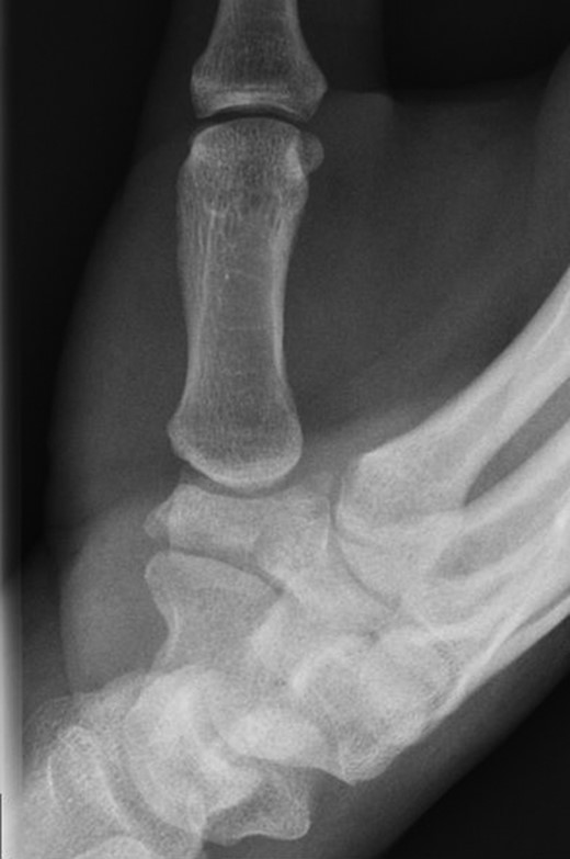
A 49-year-old man fell from his mountain bike at speed and presented to his local secondary care hospital with an injury to the base of his left thumb. There was pain and swelling at the base of the thumb with painful restriction of movements. Initial radiographs were deemed to be normal and he was treated as a soft tissue injury with a period of immobilization followed by physiotherapy. The patient was referred to us four months later as the pain and weakness were persisting. A thorough retrospective perusal of his history and imaging to date was carried out. A review of his initial plain radiographs (Fig. 1) and MRI scans indicated a fracture of the left trapezium and a CT scan was obtained to further characterize the anatomy of the fracture.

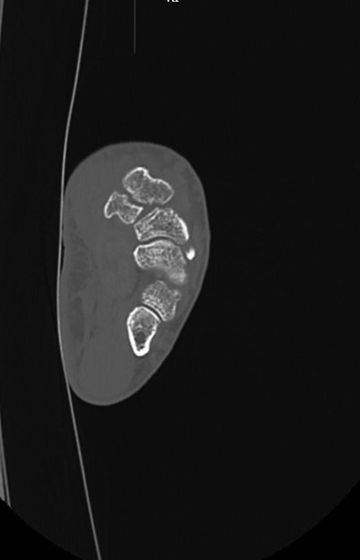
The CT scan (Fig. 2) showed a coronal fracture of the trapezium which was not united. At this stage the symptoms of pain and instability continued to persist and were preventing him returning to mountain biking. As the imaging also showed that there were no degenerative changes in either the trapezio-metacarpal joint or the scapho-trapezial joint, the decision was made to proceed with debridement of the non-union and rigid internal fixation.

The trapezium was exposed through a Wagner approach. Intra-operatively a coronal split of the trapezium was found with no evidence of union. The fracture site was debrided, reduced and fixed with two headless compression screws. The thumb was immobilized in a plaster following surgery. Six weeks following surgery plaster was removed and the patient started physiotherapy to regain range of movement and strength. His plain radiographs at this point showed progression to union.
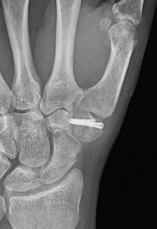
At 3 months from injury, the patient had complete resolution of his symptoms and had gone back to mountain biking. Range of movement had improved remarkably and he was pain free. Plain radiographs confirmed union across the fracture site (Fig. 3).

DISCUSSION
Isolated fractures of the trapezium are rare and isolated fractures even more so [3–6]. They occur more commonly in conjunction with fractures of the thumb metacarpal or other carpal bones [2, 4, 7]. A fall onto an outstretched hand with the wrist in radial deviation and direct commissural force with shear are reported to be the most frequent mechanisms of injury. Walker et al. [6] have classified fractures of the trapezium into five categories based on the anatomical part involved.
Owing to the distinctive shape and position of the trapezium among the other carpal bones, fractures have historically been difficult to identify and treat. Improperly positioned radiographs and overlapping of the other carpal bones make trapezium fractures difficult to delineate. A Robert’s view and Bett’s view are useful in this regard [1]. The Robert’s view is a true antero-posterior radiograph of the thumb with the forearm in maximal pronation, dorsum of the thumb resting on the cassette and the beam at 90° to the cassette. The Bett’s view is a true lateral view of the thumb ray with palm on the cassette, the hand then being pronated 15°–20° and the beam angled proximally ~15°.
MR and CT also play a vital role in the diagnosis of these injuries especially in the setting of other major injuries. The clinical symptoms mainly comprise pain, instability and weakness of grip [8, 9]. These symptoms in the absence of clear radiographic findings can often be misdiagnosed as a soft tissue injury contributing to these injuries being missed in the emergency setting.
Several techniques for treatment have been described, including closed reduction and immobilization in a plaster, percutaneous wire fixation, external fixation and open reduction with internal fixation [1, 3–6, 10].
In a non-union, the fracture needs to be opened so that it can be debrided, accurately reduced and fixed. Several authors also advocate transfixing the thumb carpometacarpal joint to neutralize forces acting on the fixed fracture during healing.
To our knowledge internal fixation for a non-union of the trapezium has not been reported. Our patient presented to us late, following treatment for a ligament sprain. Careful clinical examination and additional cross sectional imaging coupled with a high index of suspicion is essential in making a diagnosis of these injuries. If degenerative changes had been present in our case, the prognosis from fixation would have been more guarded, and consideration would have to be given to salvage procedures, such as trapeziectomy.
These are rare and challenging injuries to diagnose and can often be missed. Open reduction and internal fixation, even after delayed diagnosis and late surgery, can result in good functional outcome.
CONFLICT OF INTEREST STATEMENT
None declared.


