-
PDF
- Split View
-
Views
-
Cite
Cite
Tomislav Tokic, Iva Kirac, Davor Hrabar, Branko Troskot, Miroslav Bekavac-Beslin, Laparoscopic transhiatal resection of a large mid-esophageal diverticulum: a case report, Journal of Surgical Case Reports, Volume 2018, Issue 4, April 2018, rjy066, https://doi.org/10.1093/jscr/rjy066
Close - Share Icon Share
Abstract
This is a description of transhiatal laparoscopic approach for mid-esophageal diverticulum. Traditionally mid-esophageal diverticula are approached by thoracotomy or thoracoscopy, with the laparoscopic technique being reserved for epiphrenic diverticula. A 78-year-old Caucasian female with a secondary dilatative ischemic cardiomyopathy presented with dysphagia, tenderness in the epigastrium and a considerable weight loss. A large mid-esophageal diverticulum was found on barium swallow and confirmed by CT scan. Underlying achalasia was recorded on manometry. The patient underwent diverticulectomy via transhiatal approach, followed by Heller myotomy and Dor fundoplication. Throughout the procedure auxiliary, esophagoscopic image was provided by interventional gastroenterologist due to a very narrow operating field and lack of orientation points. Based on our experience with this case, we propose transhiatal approach as a feasible alternative to thoracoscopy, in particular with patients who suffer from cardiac or pulmonary co-morbidities which make traditional techniques of high risk.
INTRODUCTION
Thoracic esophageal diverticula are rare disorders which are treated when symptomatic, and mostly occurring in elder patients [1]. Based on their location, they are divided into epiphrenic and mid-esophageal diverticula, the line elucidating them being 5 cm below the tracheal carina [2]. Mid-esophageal diverticula are considered to be traction diverticula, and their primary cause is thought to be mediastinal inflammation when scarring or fibrous tissue pull a portion of the esophageal wall. They affect all esophageal wall layers, therefore they are ‘true’ diverticula, unlike epiphrenic diverticula which are almost always pulsion or ‘false’ diverticula, meaning that they are mucosal outpunching through a weakness in muscular layer [3]. Symptoms indicating surgical treatment, such as dysphagia, regurgitation, heartburn, halitosis and weight loss, are all results of underlying motility disorder.
Reviewing the literature, left-sided thoracotomy and thoracoscopy are common approaches for resection of thoracic esophageal diverticula [4, 5]. While thoracoscopy is preferred in treating mid-esophageal diverticula due to shorter recovery time and decreased morbidity, there is a disadvantage of great difficulty to perform a myotomy or antireflux procedure. In recent years laparoscopic transhiatal approach has become increasingly more implemented, mainly for resection of epiphrenic diverticula [6]. The laparoscopic approach provides larger mobility for performing myotomy and reflux procedures, such as Dor fundoplication. Herein, we report a modification of the application of the laparoscopic surgical approach to a mid-esophageal diverticulum.
CASE REPORT
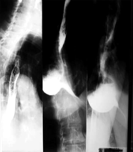
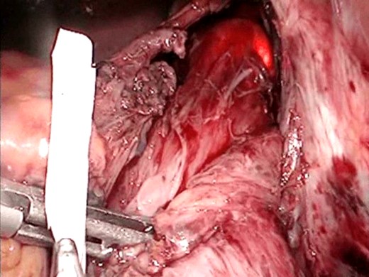
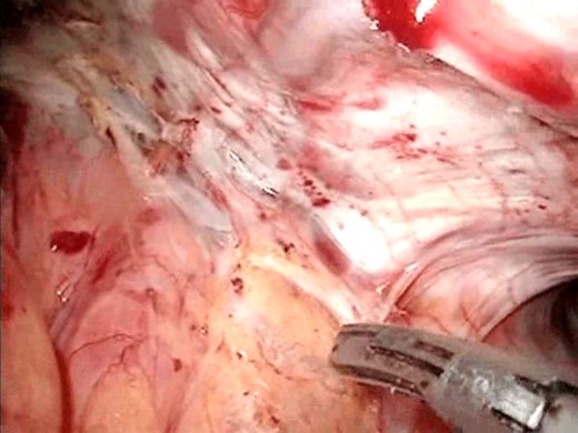
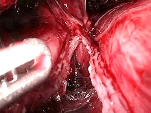
A 78-year-old Caucasian woman presented with recurrent dysphagia, slight tenderness in the epigastrium and substantial weight loss. Difficulty with solid food digestion and regurgitation were present for almost 10 years. The symptoms had exacerbated four months before intervention during which period the patient was fed through a nasogastric tube. CT scan, barium swallow (Fig. 1) and esophagogastroduodenoscopy showed a large mid-esophageal diverticulum. The neck of diverticulum was within 5 cm of the carina and was 4 cm wide. The diameter of the sac reached 10 cm, and narrow esophagogastric junction suggested achalasia, which was confirmed by manometry. The patient suffered a heart attack 3 years ago and has secondary dilatative ischemic cardiomyopathy. Due to cardiac co-morbidities, surgical intervention was graded as a high-risk procedure (ASA III), with patient’s quality of life being crucial in decision making. The patient was placed in supine position in reverse Trendelenburg with five ports placed as earlier described for transhiatal epiphrenic diverticula operations [7]. Throughout the procedure the esophagoscopic image was provided by an interventional gastroenterologist and dissection was performed under the guidance of both laparoscopic and endoscopic image (Fig. 2). This was essential due to a very narrow operating field and a lack of orientation points. The liver was retracted, and crura of the diaphragm were dissected with Ultracision (Ethicon Endo-Surgery Inc., Cincinnati, OH, USA) (Fig. 3), after which mediastinum was entered. The esophagus was bluntly dissected around its entire circumference and the gastroesophageal junction was encircled with an umbilical tape to ensure easier traction. The diverticulum was approached by both blunt dissection and electrocauterization. Resection of the wide neck of diverticulum was done with EndoGIA (Covidien, Mansfield, MA, USA) (Fig. 4). Another cartridge was used for dissection of the superior portion of the diverticulum. Possible air leakage was tested by irrigation of the field and was negative. Dissected diverticulum was withdrawn from the mediastinum and placed into an endobag. Heller myotomy was performed 5 cm orally and 2 cm distally from the gastroesophageal junction for reduction of achalasia. After the cruroplasty had been performed, the anterior fundus wall was sutured to the muscular edges of myotomy (Dor fundoplication) to prevent possible postoperative complications, such as gastroesophageal reflux disease (GERD). Operating time was 6 h, and the procedure went without any complications.

Preoperative barium swallow. The preoperative barium swallow images showing a 10 × 8 cm diverticulum whose 4 cm wide neck was situated ~5 cm from the carina.

Gastroscopic guidance. This was needed for orientation within the mediastinum.


Resecting the diverticulum. The diverticulum was resected with a gastrointestinal stapler.
Surgically, patient’s recovery was uneventful; however, patient suffered from an acute coronary incident. That was the reason for prolongation of the Intensive Care Unit (ICU) stay to 5 days. Water soluble contrast was administered after which per oral nutrition was initiated. On Day 5 of ICU stay, the patient was transferred to cardiology open ward.
On 1 and 6 months postoperative follow-up barium swallow demonstrated clear passage. The patient had no complaints and was gaining weight.
DISCUSSION
Since the first description of esophageal diverticula by Ludlow in 1769 and by Zenker and von Ziemssen in 1874 numerous surgical techniques have been described amongst which thoracotomy and lately thoracoscopy are preferred. Left thoracotomy was a preferred technique as it allows an easy approach to the thoracic diverticulum. However, due to high morbidity and mortality [4], thoracoscopy was favored, and still is a standard procedure regarding mid-esophageal diverticula [8]. A severe disadvantage of the thoracoscopic approach is a lack of mobility when performing myotomy and antireflux procedures, which have become standard complementary procedures accompanying diverticulectomy.
Recently, many authors report the laparoscopic transhiatal approach to epiphrenic diverticula as a satisfying alternative to thoracoscopy, both single port [9] and multiport [7, 10] approach being described. The most significant benefit of this method is that it allows surgical resection parallel to and along with esophagus versus perpendicular in thoracoscopic approach, enabling easier stapler manipulation. Another argument for this approach is the possibility of myotomy or antireflux procedure, which are performed for underlying neuromuscular dysfunction and prevention of GERD, respectively. This approach is performed only on epiphrenic diverticula and reviewing the literature only one case of mid-esophageal resection was found [11], although on a smaller size diverticulum.
As in our case, the aforementioned authors report a patient with severe co-morbidities, which required an approach previously reserved for lower esophageal diverticula. We concur with their findings and based on our experience propose the extension of already established technique for lower esophageal diverticula to mid-esophageal diverticula.
Based on our experience, we suggest the transhiatal laparoscopic approach to mid-esophageal diverticulum as a feasible alternative to thoracoscopy, in particular when patient presents with severe cardiac or lung co-morbidities which make the thoracic approach of exceptionally high risk.
ACKNOWLEDGEMENTS
Filip Karabelj for technical assistance.
CONFLICT OF INTEREST STATEMENT
The author(s) declare that they have no competing interests.
FUNDING
No funding was obtained for this study.
AUTHORS’ CONTRIBUTION
T.T. wrote the article and did the literature search. I.K. edited the video and wrote the article. D.H. and B.T. performed gastroscopy during the procedure and provided constructive critics to the article. M.B.B. performed the procedure and structured the article.
REFERENCES
- esophageal achalasia
- computed tomography
- lung
- ischemic cardiomyopathy
- barium swallow
- weight reduction
- deglutition disorders
- esophagomyotomy
- diverticulum
- laparoscopy
- manometry
- thoracoscopy
- thoracotomy
- european continental ancestry group
- heart
- morbidity
- acquired supradiaphragmatic diverticulum of esophagus
- diverticulectomy
- dor fundoplication
- epigastrium
- gastroenterologists


