-
PDF
- Split View
-
Views
-
Cite
Cite
J van Bastelaar, L M van Roozendaal, M Meesters-Caberg, Surgical removal of fibrous axillary seroma pocket and closing of dead space using a lattisimus dorsi flap, Journal of Surgical Case Reports, Volume 2018, Issue 3, March 2018, rjy032, https://doi.org/10.1093/jscr/rjy032
Close - Share Icon Share
Abstract
Seroma formation after axillary dissection is a common problem in breast cancer surgery. We report the case of a 68-year-old female with breast cancer who underwent a wide local excision and axillary clearance due to stage III breast cancer. Patient received post-operative whole breast irradiation therapy and developed a painful, infected seroma one month after surgery. This was treated with antibiotic therapy after which the infection subsided. One year after surgery patient presented with a painful persisting seroma in the left axilla. We decided to surgically treat the seroma by removing the fibrous seroma capsula and closing of the dead space with a latissimus dorsi flap. Six weeks after surgery, patient was pain and seroma free and was happy with the surgical result. Latissimus dorsi flap harvesting is an ideal way to treat persisting fibrous encapsulated seroma pockets after axillary clearance in the treatment of breast cancer.
INTRODUCTION
Seroma formation after breast cancer surgery is defined as a collection of serous fluid containing blood plasma and/or lymph fluid under the skin flaps or in the axilla. The reported incidence of seroma varies greatly, ranging from 3% to more than 90% [1–3]. Seroma formation can lead to patient discomfort, repeated seroma aspirations with the risk of infection, prolonged hospital stay, delayed wound healing, skin flap necrosis, delay in commencing adjuvant therapies and higher surgical expenditures [2, 4, 5].
The pathophysiology of seroma formation has been extensively analysed. The extent of axillary lymph node involvement, type and extent of breast surgery and the use of electrocautery have all been related to seroma formation. In recent years there have been many publications on effective techniques to prevent seroma formation. These techniques all appear to have one common denominator: reduction of the dead space [6]. There are however very few publications on (surgical) treatment of fibrous encapsulated seromas. Most articles are case reports, none of which describe filling of the dead space with flap harvesting [7, 8].
CASE PRESENTATION
A 68-year-old female was referred to our breast clinic by her primary care physician after undergoing screening mammography. Upon mammography, a breast mass was visualized in the left upper quadrant, measuring 13 mm × 8 mm. Ultrasonography of the left axilla revealed two suspicious lymph nodes with cortical thickening. The breast mass and a suspicious lymph node were biopsied. Pathology revealed a hormone positive ductal breast carcinoma and a lymph node metastasis in the left axilla. Patient was planned for wide local excision and axillary clearance under general anaesthesia. Routine pathology examination revealed a 1.1 cm grade 1 ductal carcinoma with clear resection margins and 18 lymph nodes, one containing a macro metastasis (pT1cN1 (1/18)). Patient was discussed postoperatively in the multidisciplinary breast cancer meeting and adjuvant Tamoxifen and breast radiotherapy were instituted (16 fractions in a total dose of 42.56 Gy).
One month after surgery patient presented to our breast clinic with a fever and swelling and redness of the skin in the left axilla. Under suspicion of an infected seroma, seroma aspiration was performed and sent for culture. Antibiotic treatment was instituted for one week and she remained free of symptoms for a couple of months.
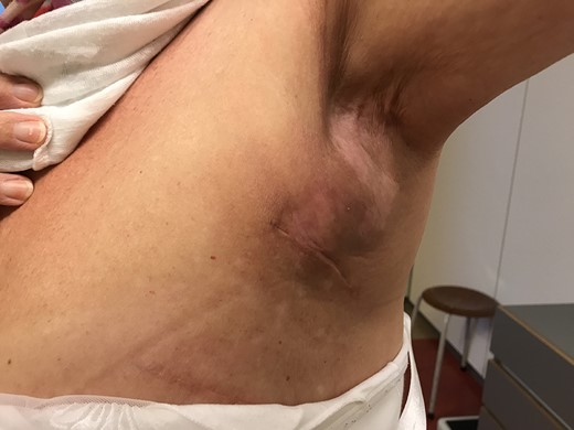
One year after surgery and radiotherapy, our patient was seen in the breast clinic for follow up. Mammography was unremarkable. Upon physical examination there was an apparent fibrotic seroma pocket that had persisted (Fig. 1). The overlying skin was sensitive to touch and displayed radiation dermatitis. Her arm and shoulder function were impaired. Range of motion was limited to 110° when abducting her arm.

Fibrous seroma capsula of the left axilla after axillary clearance and radiotherapy.
This case was discussed with our reconstructive breast team and after informed consent was obtained a surgical excision of the fibrous seroma pocket and the overlying skin was performed. The dead space was closed with a latissimus dorsi skin and muscle flap. Low suction drains were left in place. The drains were removed on the second day post-surgery.
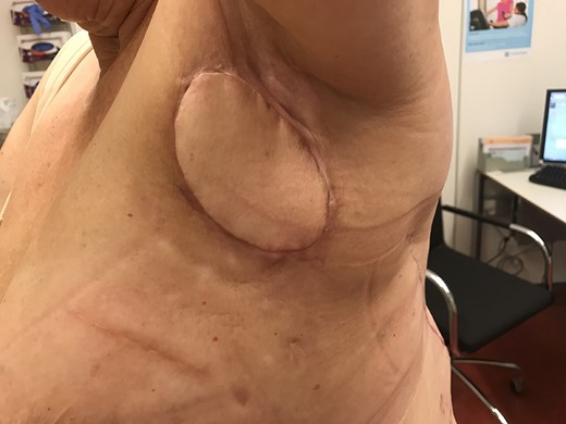
Six weeks after surgery patient was evaluated in the outpatient clinic. The pain had subsided and there was no clinical seroma. Movement of her shoulder had improved greatly and abduction of the right arm was now possible up to 160° (Fig. 2).

After surgical excision of seroma pocket and filling of the dead space with a latissimus dorsi skin-muscle flap.
DISCUSSION
Seroma formation occurs frequently after mastectomy or axillary clearance in breast cancer surgery. Seroma has significant impact on patients’ quality of life and could delay the initiation of adjuvant treatment. It is well known that seroma occurs and causes complaints in the early weeks post-surgery and/or radiotherapy. In the literature, a great number of articles can be found on seroma formation, its risk factors, consequences, and early treatment. Reported treatment options are limited, mostly consisting of repeated aspirations and treatment of infection where necessary.
Many factors are held responsible (surgical technique, instruments used for dissection, obliterating the dead space, use of drains) causing seroma formation. The key to reducing seroma formation and its sequelae seems to lie in reduction of the dead space [9]. Previous retrospective studies have proven that reduction of the dead space after mastectomy is beneficial in seroma formation and that it furthermore reduces complications associated with seroma formation. There are no studies to date describing the surgical treatment of fibrous encapsulated seromas after axillary clearance. This case demonstrates that filling the dead space with skin/muscle flap harvesting is an ideal technique to combat seroma formation. As seen in many studies analysing flap fixation after mastectomy, closing of the axilla can be troublesome. As published by Van Bemmel et al. in a systematic review, closing of the axillary dead space leads to reduction of seroma formation. Ten Wolde et al. retrospectively evaluated 27 patients undergoing axillary lymph node dissection with and without quilting. In the non-quilting group, seroma was present in 75% and in the quilting group 36.4%. There were however no significant differences in number of seroma aspirations [10].
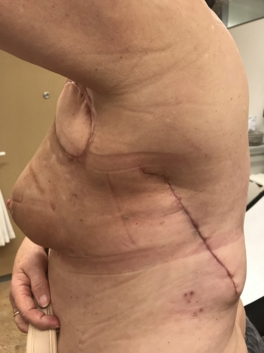
Given the large burden that this is causing to patients and the varying chance of success, an important role is dedicated to preventing seroma formation. We expect that the rate of chronic seroma is underestimated and generates high rates of local discomfort, and limitations in shoulder function, especially in axillary seroma. In the literature, some refer to treating persisting seroma by reinserting drains, talcage or surgical intervention and application of the quilting technique. Since the seroma pocket is covered with fibrous tissue, these techniques are inadequate in chronic seroma. Collaboration with a reconstructive surgeon therefore seems favourable in order to remove the fibrous capsule and close the dead space with a latissimus dorsi flap (Fig. 3).

Lateral view after surgical excision of seroma pocket and filling of the dead space with a latissimus dorsi skin-muscle flap.
LEARNING POINTS
Reducing the dead space is pivotal in preventing seroma formation.
Little is known about the best way to treat encapsulated seroma pockets in the axilla.
Removal of encapsulated axillary seroma pockets and closing the dead space with latissimus dorsi flap seems to be a successful surgical procedure.
CONFLICT OF INTEREST STATEMENT
None declared.


