-
PDF
- Split View
-
Views
-
Cite
Cite
Julian J L Leow, Jason J F Fleming, Richard R O Oakley, A case series on the conservative management of the bony skull in patients with aggressive skin carcinomas of the scalp, Journal of Surgical Case Reports, Volume 2018, Issue 12, December 2018, rjy326, https://doi.org/10.1093/jscr/rjy326
Close - Share Icon Share
Abstract
Few studies examine techniques of surgical resection for scalp malignancies to ensure clear margins. We present a case series utilizing outer cortex removal in patients without evidence of bony or pericranial invasion. A retrospective casenote review is presented of three cases treated in a tertiary Head and Neck Cancer Centre. An outer table removal approach was utilized based on the absence of bony involvement either on pre-operative imaging or from intra-operative findings. All cases underwent an outer table drilldown procedure. Tumour histology included high grade carcinoma of unknown origin, malignant cylindroma and squamous cell carcinoma. Complete excisions with adequate deep margins were achieved in 100% cases. Overall disease-free survival was 66.6% and local control rate was 100%. This technique allows a high degree of local control, notably at the deep margin. There is little morbidity and it avoids the complications associated with full thickness calvarial resection.
INTRODUCTION
There is a paucity of published evidence for the management of malignancies in the scalp. Primary scalp malignancy is rare but can occur in all five layers of the scalp, most often the skin. Invasive scalp malignancies can rapidly spread through the layers of the scalp and involve the calvarial bone or invade through it resulting in intracranial extension.
We present a small case series utilizing outer cortex removal in carefully selected patients without evidence of bony or pericranial invasion.
CASE SERIES
Three cases treated in a tertiary Head and Neck Cancer Centre were identified between July 2014 and April 2015 (Table 1). All patients were discussed and management planned at the Multi-Disciplinary Team meeting. All patients had confirmed malignant disease based on punch/incisional biopsies and disease management was planned based on pathological and radiological features. An outer table removal approach was decided based on the absence of bony involvement on pre-operative imaging, with the plan for an intra-operative assessment for any macroscopic bony invasion. The limitations in sensitivity of radiological investigations to assess bony involvement and pathological definitions for disease clearance in the presence of a bony margin often contribute to documented inadequate deep excision margins.
| Case 1 . | Case 2 . | Case 3 . |
|---|---|---|
| Diagnosis | Diagnosis | Diagnosis |
| High grade carcinoma with 4 cm left level V neck lump | Low grade malignant cylindroma | Recurrent well-differentiated squamous cell carcinoma |
|
|
|
| Case 1 . | Case 2 . | Case 3 . |
|---|---|---|
| Diagnosis | Diagnosis | Diagnosis |
| High grade carcinoma with 4 cm left level V neck lump | Low grade malignant cylindroma | Recurrent well-differentiated squamous cell carcinoma |
|
|
|
| Case 1 . | Case 2 . | Case 3 . |
|---|---|---|
| Diagnosis | Diagnosis | Diagnosis |
| High grade carcinoma with 4 cm left level V neck lump | Low grade malignant cylindroma | Recurrent well-differentiated squamous cell carcinoma |
|
|
|
| Case 1 . | Case 2 . | Case 3 . |
|---|---|---|
| Diagnosis | Diagnosis | Diagnosis |
| High grade carcinoma with 4 cm left level V neck lump | Low grade malignant cylindroma | Recurrent well-differentiated squamous cell carcinoma |
|
|
|
Surgical technique
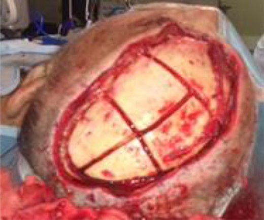
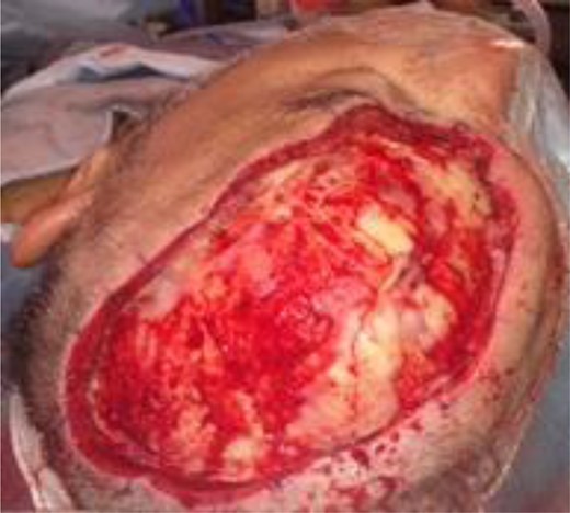
Each scalp lesion was excised whole with a 6 mm–2 cm excision margin as directed by the pathological diagnosis, leaving the pericranium attached to bone. Marginal biopsies were sent for frozen section analysis. The exposed periosteum was then stripped from the underlying skull. A bony margin corresponding with the overlying excision was drilled with a Rosehead burr to bevel the margins on the outer cortical plate down to the diploic space—identified by characteristic bleeding. The encircled outer cortex was then divided into sections with the drill (Fig. 1), and each section was removed with a mallet and osteotome leaving the inner cortex intact (Fig. 2). Osteotomes should be introduced at an angle to reduce the risk of intracranial penetration. All sections were sent for final histological analysis. The tissue reconstruction utilized in each case is summarized in Table 2, and should be tailored to the individual defect and patient factors.


Surgery performed. A variety of techniques are used to ensure adequate coverage of the inner cortical plate
| Case 1 . | Case 2 . | Case 3 . |
|---|---|---|
|
|
|
| Case 1 . | Case 2 . | Case 3 . |
|---|---|---|
|
|
|
Surgery performed. A variety of techniques are used to ensure adequate coverage of the inner cortical plate
| Case 1 . | Case 2 . | Case 3 . |
|---|---|---|
|
|
|
| Case 1 . | Case 2 . | Case 3 . |
|---|---|---|
|
|
|
Results and analysis
Three cases underwent an outer table drilldown procedure during the study time period. Tumour histology included high grade carcinoma of unknown origin, malignant cylindroma and squamous cell carcinoma (SCC). Disease-free survival was 66.6% (two patients) with a local control rate of 100% (three patients). Complete excision with clear deep margins were achieved in 100% (three patients), negating the need for further adjuvant treatment and therefore limiting morbidity. Of note, subsequent histological analysis revealed extracapsular spread in more than one of the excised lymph nodes from a concurrent neck dissection in the high-grade carcinoma patient and on follow up, new metastatic disease was discovered. Local control however was maintained.
DISCUSSION
There are no current guidelines on the depth of surgical excision for scalp malignancies. The concept for this surgical approach is translated from SCC of the mandible where partial thickness mandibulectomy is acceptable in certain circumstances, if the outer table of the cortical bone is not directly invaded [1].
Here we present three cases where the outer table of the calvarium was removed to ensure an oncologically complete resection in the absence of bony invasion. Removal of the outer table of the calvarium is suggested as it increases the surgical margin whilst causing minimal morbidity and risk. Although the risk is low, there is risk of penetration of the inner table. The larger the defect, the greater the convexity of the skull which increases the risk of perforation of the inner table and dural tear [2]. Dividing the defect into smaller sections before removal reduces the risk of perforation [3]. Other risks include cerebral spinal fluid leak and meningitis after dural tear. Perforation of the sagittal sinus is also a risk when operating around the sagittal midline. Variable skull thickness depending on site and patient factors should be taken into account.
The periosteum and the underlying skull should be examined clinically for signs of invasion. Early signs of outer cortex invasion are bone stippling and difficulty in peeling off the periosteum [4]. If no signs of clinical invasion are present in the periosteum and the skull, then only the outer table of the calvaria should be removed.
Removing outer table
Different instruments have been reported including straight and curved osteotomes, reciprocating saws, oscillating saw and Gigli saws. When utilizing osteotomes, it’s important to have sharp contact edges and the correct angulation. To minimize the impact of the curvature of the skull and the potential breach of the inner cortex, margins of the outer cortical plate should be bevelled down to the diploic space to allow access of the osteotomes.
Diagnostic dilemma
A diagnostic dilemma exists between the certainty of diagnostic image techniques and incidence of microscopic or macroscopic pericranial or cranial invasion at the deep margin of scalp tumours. For example, Hong et al. [5] reported an average 3.4 mm underestimation of the extent of SCC invasion into the mandible when using radiological imaging. For head and neck cancer detection, reported sensitivity values for positron emission tomography/computed tomography (CT) scans range from 0.68% [6], for CT scans—85.7% [7] and magnetic resonance imaging scans—75.8% [8].
Deep margins
There have been several reports of incomplete excisions particularly involving the deep margins. Khan et al. [9] found that from their cohort of 633 patients with scalp SCC, 94% of (45/48) patients with incomplete excision had incomplete excision at the deep margins. The authors recommended deeper excision of the tumour past macroscopic clinically clear deep planes. The study by Bovill and Banwell [10] also reported that incompletely excised lesions frequently involved the deep margins of the tumour.
CONCLUSION
Removal of the outer table of the calvarium allows a high degree of local control in scalp malignancy with improved likelihood of clear deep margins. This procedure has little morbidity and avoids the complications associated with complete calvarial excision. In selected patients, outer table drilldown offers a safe, oncologically sound approach for scalp malignancies.
ACKNOWLEDGEMENTS
No other contributors. This article was presented as a poster at the scientific meeting of the British Association of Head and Neck Oncologists (BAHNO).
CONFLICT OF INTEREST STATEMENT
None declared.
FUNDING
None.


