-
PDF
- Split View
-
Views
-
Cite
Cite
Lauryn A Ullrich, William Streiff, David R Mariner, Boyoung Song, Melissa A Obmann, Shivprasad D Nikam, Non-operative management of isolated spontaneous superior mesenteric artery dissection, Journal of Surgical Case Reports, Volume 2018, Issue 10, October 2018, rjy274, https://doi.org/10.1093/jscr/rjy274
Close - Share Icon Share
Abstract
Isolated spontaneous superior mesenteric artery (SMA) dissection is a rare differential for patients presenting with abdominal pain. Due to limited cases reported, management strategies have been poorly defined. We present the case of a 49-year-old male with history of hypertension and ischemic colitis, presenting with abdominal pain. CT imaging demonstrated a thrombosed dissection of the SMA extending into second and third order braches. He was managed conservatively with therapeutic anticoagulation. His symptoms improved and upon discharge he was transitioned to aspirin and warfarin. Repeat CT imaging continued to show the dissection with resolution of the SMA thrombus. Spontaneous SMA dissection is exceedingly rare with no universally agreed upon standard of care for treatment. Operative intervention should be reserved for failed conservative management or vascular compromise. Understanding the current treatment options helps ensure a favorable patient outcome.
INTRODUCTION
Isolated spontaneous superior mesenteric artery (SMA) dissection is a rarely reported and potentially fatal cause of acute abdominal pain. In a MEDLINE literature search in 2011 only 168 cases were reported with an estimated incidence of 0.06% in post-mortem studies [1, 2]. Clinical presentation of SMA spontaneous dissection has been found to most commonly occur in males in their mid-50s presenting with acute epigastric pain usually after overeating and drinking [1, 3]. Of the cases reported, uncontrolled hypertension accounts for 30% of coexisting medical conditions followed by smoking, intra-abdominal cancer and hypercholesterolemia [1, 2]. In contrast, there is a low correlation with atherosclerosis and atherosclerotic heart disease [1, 3].
Due to the rarity of this disease process the underlying natural course of spontaneous SMA dissection has yet to be determined [1, 3]. The SMA is the second most frequent peripheral artery to be affected by dissection following the internal carotid artery [4]. An accepted theory to support spontaneous dissection is based on the anatomy and the hemodynamic force created by the blood flowing through the SMA. It is believed that the convex curvature of the artery leads to dissection occurring in proximity to the ostium [2, 5]. Interestingly, there has been a positive correlation found between pain severity and the length of dissection on imaging [6].
Due to the small number of cases, management strategies in the past have been poorly defined and both operative and non-operative approaches have been employed. We present to you a case presentation of spontaneous SMA dissection that was managed non-operatively with anticoagulation.
CASE REPORT
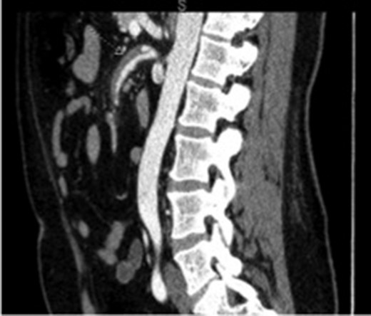
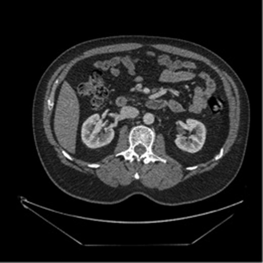
The patient was a 49-year-old male with history of hypertension presenting for evaluation for sudden onset of bilateral lower quadrant abdominal pain, nausea, emesis and non-bloody diarrhea starting 4 h prior to presentation. The patient denied prior abdominal surgeries, tobacco or illicit drug use. His history included hospitalization a year earlier for painless rectal bleeding, concerning for ischemic colitis, which was managed non-operatively. Current medications included Aspirin 81 mg and Valsartan-HCTZ. Physical exam revealed only mild left lower quadrant tenderness, and the labs values were quite unremarkable. A CT scan of the abdomen and pelvis with intravenous contrast demonstrated a thrombosed dissection, originating 2 cm from the ostium of the SMA with extension into the second and third order branches (Figs 1 and 2).

Initial imaging showing a thrombosed dissection, originating 2 cm from the ostium of the SMA with extension into the second and third order branches.

The patient was admitted for observation, non-operative management and intravenous hydration, along with bowel rest with therapeutic anticoagulation. His abdominal pain improved and he was discharged home on hospital Day 5 on aspirin and warfarin.
Repeat CT imaging 3 months after this hospitalization continued to show the SMA dissection with resolution of the SMA thrombus (Fig. 2). His warfarin was discontinued in exchange for a dual antiplatelet therapy.
DISCUSSION
Spontaneous SMA dissection is exceedingly rare with no universally agreed upon standard of care for treatment. Current literature recommends that clinical condition and imaging findings should guide management; however, non-operative management with or without anticoagulation has been reported to be successful in patients that are both asymptomatic and symptomatic without evidence of bowel ischemia [1, 6–8]. The rationale of medical therapy is to prevent further hematoma of the stenotic arterial segment and to prevent distal embolization [8]. Anticoagulants are utilized to prevent clot and emboli formation which helps to recanalize the true lumen in comparison to antiplatelet therapy that prevents arterial thrombosis. Anticoagulant and antiplatelet therapy duration and dosing remain debatable.
In symptomatic patients undergoing conservative therapy there are reports of resolution of abdominal pain in 7–14 days. Current recommendations are to continue anticoagulation or antiplatelet therapy until there is no dissection progression or until there is complete resolution of the dissection on imaging [6]. In regards to our patient, antiplatelet and anticoagulation were used successfully without progression of the patient’s SMA dissection. There have been cases reported that did not use anticoagulant or antiplatelet therapies that showed no progression of the dissection.
Surgical management remains a mainstay of treatment for failed conservative management, vascular compromise or an acute abdomen. Endovascular treatments should be considered with use of a flow diverting stent in patients with suspected intestinal ischemia, angina abdominis, worsening abdominal pain despite conservative management, true lumen compression >80%, SMA aneurysm of >2 cm or dissection progression [1, 2, 4, 8]. For patients presenting with evidence of advanced intestinal ischemia or aneurysmal rupture, emergent open surgical intervention is indicated such as SMA re-implantation, resection or repair of the affected segment with interposition bypass or graft [1, 2, 8].
With the limited amount of literature available concerning the management and treatment of spontaneous SMA dissection, this case further highlights the successful use of non-operative management in combination with anticoagulation for the treatment of spontaneous SMA dissection. A good understanding of the current literature, available treatment options and judicious use of open and endovascular management helps ensure a favorable patient outcome.
Conflict of Interest statement
None declared.


