


Abstract
Hemangioma is the most common benign hepatic tumor. We present the case of a patient with a giant symptomatic hemangioma, treated with segmental liver resection using the Da Vinci Robotic System. A 38-year-old woman presented to our surgical out-patient Department complaining about abdominal discomfort and recurrent episodes of acute abdominal pain. CT-scan and MRI imaging of the abdomen revealed the presence of a giant hepatic hemangioma (>5 cm) involving segments VI and VII. Robotic right segmental hepatectomy was performed. The procedure was successfully completed in 120 min and with intraoperative blood loss of only 450 ml. Postoperative period was uneventful and the patient was discharged on the second postoperative day. In case of giant hemangiomas, a minimally invasive robotic major hepatic resection is a viable option that can be performed with minimal complications. A careful preoperative and intraoperative strategy is required, while significant experience in liver and robotic surgery is mandatory.
INTRODUCTION
Minimally invasive hepatic surgery has shown a dramatic development during the last decade, progressing from minor hepatic interventions to major hepatectomies with outcomes comparable to open interventions [1]. This technique was initially developed to avoid disadvantages of conventional laparoscopic surgery.
Robotic surgery of the liver can enhance the surgeon's laparoscopic skills through a magnified 3D view and instruments with seven degrees of freedom compared to conventional laparoscopy [2]. Therefore, hepatic surgery can be realized with precision, minimal acts, smaller incisions, decreased blood loss, less pain and quicker healing time. Moreover, also reduction of length of hospital stay, transfusions and analgetics is achieved [3].
Robotic surgery for the treatment of giant hepatic hemangiomas is currently very limited; however, it can present favorable results. We report the case of a robotic right segmental hepatectomy for a giant symptomatic hemangioma in a young female patient, which led to successful treatment.
CASE REPORT
A 38-year-old woman was referred to the surgical out-patient Department of our Center presenting abdominal discomfort and recurrent episodes of acute abdominal pain during the previous 6 months. Her clinical and surgical history were free, and the patient did not mention any other symptoms. Physical examination only revealed tenderness during palpation of the right upper abdominal quadrant. Laboratory blood examination showed results within normal limits. CT-scan and MRI were ordered, in order to further investigate this condition. These imaging controls revealed the presence of a giant hemangioma (~9.6 cm in diameter), involving segments VI and VII of the liver (Figs 1 and 2). Therefore, in this symptomatic patient, considering the location, size and possible future complications of the lesion, a right segmental hepatectomy was decided.
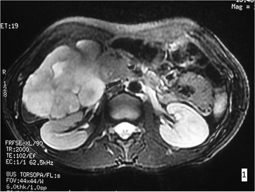
MRI imaging. In the right hepatic lobe a lesion of 9.6 × 8.9 × 7.9 cm3 is observed, hyperdense in T2W presenting contrast enhancement.
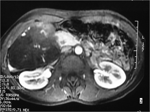
MRI imaging. Involvement of hepatic segments VI and VII.
The procedure was carried out under general anaesthesia and the patient was monitored by ECG. Peripheral oxygen saturation (SpO2), end-tidal CO2 concentration and body temperature were measured throughout the whole operation. Gastric intubation and bladder catheterization were realized. A central venous line was placed and an intra-arterial catheter was positioned for continuous monitoring of the blood pressure and gas analysis. The patient was placed in supine position with parted legs in 20° Trendelenburg position. Pneumoperitoneum was achieved. A port was placed for the robotic camera and three additional ports were introduced. Cholecystectomy was first performed leading to a better optimal exposure of the hepatic hilum. Using the da Vinci surgical system, a right segmental hepatectomy was performed (Figs 3 and 4).
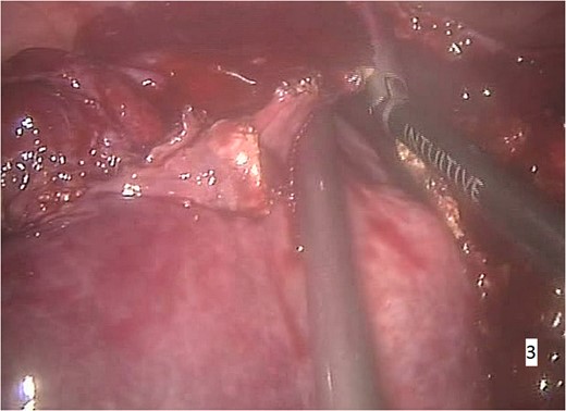
Intraoperative imaging. Segmental right hepatectomy and removal of the excised specimen through a small abdominal incision.
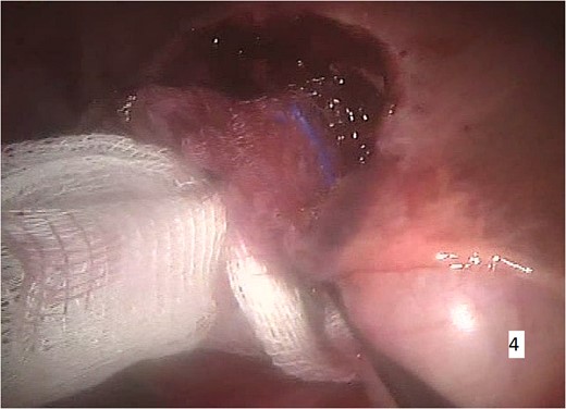
Intraoperative imaging. Minimal intraoperative blood loss.
Through this method, the procedure was completed with an operating time of 120 min; the intraoperative blood loss was only 450 ml. The patient was transferred to the intensive care unit and then to her ward. No events emerged after the surgery and the patient was discharged on the second postoperative day. Pathologic examination confirmed that the lesion was a cavernous hepatic hemangioma. On follow-up control, three months later, she reported a complete relief of her symptoms and total satisfaction for the aesthetic results.
DISCUSSION
Ηemangioma is a benign tumor of endothelial cells, characterized by an increased number of normal or abnormal vessels. Histologically, it is usually classified as capillary or cavernous. The cavernous hemangioma, as in our case, consists of large dilated blood vessels, lined by a single layer of endothelial cells [4]. It is the most common benign hepatic neoplasm, with an incidence of ~2%, and is more frequently diagnosed in women (female-to-male ratio: 4–6:1). Hemangiomas are usually small (1–2 cm in diameter), but they can be several centimeters in diameter or even larger. Giant hemangiomas measure more than 5 cm in diameter [5] and may contain areas of central necrosis or liquefaction, hemorrhage, peripheral calcification, fibrosis and thrombosis.
The vast majority of hemangiomas of the liver never cause symptoms or health problems, whereas they are discovered incidentally at the time of imaging for other medical problems, most commonly with ultrasound or abdominal CT-scan. Very large hemangiomas can cause symptoms, especially if they are located near other organs. Pain, enlargement of the liver and abdominal fullness can occur. Rarely, larger hemangiomas can rupture, causing severe pain and abdominal hemorrhage, that may become even life threatening [6]. This complication is typically encountered after trauma or biopsy.
Hepatic hemangiomas warrant therapy if they are causing significant symptoms. Surgical techniques involve open, laparoscopic or robotic resection or enuclation, while other forms, such as transplantation, ablation, embolization, radiotherapy and chemotherapy, have also been used. Reports of robotic treatment of hemangiomas are rather limited, and present experience may not be adequate in order to consider it as a first option of surgical treatment [5]. In our case, the lesion was ~5 cm in diameter and the patient suffered from disturbing symptoms, which altered her life quality. A right hepatectomy was the best choice of treatment, considering the size and the location of the hemangioma, and a minimally invasive approach via robotic techniques was chosen. The use of the Da Vinci Surgical System, as in our case, presents several technical advantages, facilitating the surgical team and improving the feasibility of a minimally invasive major liver resection [7].
During this right segmental hepatectomy, all movements of the camera and robotic instruments were precisely performed in real-time by the surgeon using ergonomic finger controls, transforming the procedure into a microsurgical approach [8]. This technique seems safe and feasible, with less complications and mortality compared to laparoscopic or open resection [9], and can also be safely used in the case of hepatic hemangiomas, as in our case no postoperative complications were present. Current reports, though, of cases of giant hepatic hemangiomas, are still limited, even considering big series of robotic hepatic resections [3, 7, 10]. Research on efficacy, cost-effectiveness and long-term oncologic effectiveness, though, is still needed.
CONCLUSION
Minimally invasive surgery has a key role in major hepatic resections at experienced centers. This technique may represent a future approach to major hepatic surgery, offering the patients significant advantages. We consider that, in experienced centers in both hepatic and robotic surgery, robotic segmental hepatectomy for the treatment of giant hemangiomas is feasible and safe, offering a series of advantages to both surgeon and patient.
CONFLICT OF INTEREST STATEMENT
None declared.
