


Abstract
Right diaphragmatic hernia is an uncommon injury following abdominal trauma. A case of delayed right post-traumatic diaphragmatic hernia is presented. The patient referred us with wheezing and cough since 1 month. A chest-abdominal computed tomography scan demonstrated a large diaphragmatic defect with liver and intestinal dislocation. The patient underwent surgical intervention with diaphragmatic repair. No complications were observed during admission and follow-up is actually negative for recurrence.
INTRODUCTION
Diaphragmatic hernia is a rare consequence of thoraco-abdominal trauma. The abdominal organ herniation trough the right diaphragm is even rarer due to the liver protective function [1].
The high morbidity and mortality of this condition require early diagnosis and rapid treatment. The case reported concerns about a patient suffering from massive delayed right diaphragmatic hernia with right liver and bowel dislocation.
CASE PRESENTATION
A 41-year-old patient was referred to our Emergency Department with complaints of wheezing and cough since 1 month. During a previous admission, a diagnosis of right basal pneumonia was done.
His medical history was significant for motorcycle accident ~20 years before involving and bladder rupture. Thoracic examination revealed decreased breathing sound and bowel sound in the right lower hemithorax.
A chest X-ray revealed right basal consolidation with inhomogenous opacity at the medium and lower chest area.
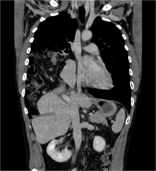
Thorax and abdominal CT showing the dislocation of right upper abdominal viscera
He was then admitted to the surgery department for a laparoscopic exploration that confirmed the radiological finding of inveterate right diaphragmatic hernia with an 8 cm defect. Because of the impossibility to reduce the liver in abdomen, due to the thoracic adhesions, a right anterolateral thoracotomy was then performed. The liver was uneventfully reinstated and colon and small bowel were replaced in anatomical position. The defect was repaired by dual mesh patch (15 × 25 cm). The postoperative course was uneventfully. Postoperative oxygen saturation was normal and a chest X-ray was performed before discharge and revealed complete re-expansion of the lung. The patient was discharged on the ninth postoperative day. The follow-up is negative for signs of recurrence after 2 years.
DISCUSSION
The diaphragmatic hernia is the herniation of abdominal organs into the chest through a diaphragmatic defect. These can be congenital or acquired [2].
Acquired diaphragmatic hernia occurs, in most of the cases, as a result of blunt or penetrating thoraco-abdominal trauma. The likelihood of occurrence of diaphragmatic hernia may be ~5% following high impact trauma [3]. Left hemidiphragmatic hernia is more common because liver exerts a protective function against the herniation of the viscera [1].
Herniation of the abdominal organs may be completely asymptomatic; due to this reason, ~66% of diaphragmatic rupture are not recognized at the time of trauma. The chest negative pressure causes the gradual migration of abdominal contents leading to the onset of symptoms [4]. We can classify this clinical condition in two types: Type I (early) and Type II (delayed). Dislocation of abdominal organs is more common in Type II hernia [1].
Clinical presentation includes gastrointestinal symptoms (abdominal pain, nausea, vomiting and sub-occlusion), respiratory (dyspnea, orthopnea and chest pain) or cardiocirculatory (hemodynamic compromission) [1–4].
The initial diagnostic tool is chest or abdominal X-ray but CT scan is the best modality to assess the extent of dislocation, the size of diaphragmatic defect and the belt-like constriction of abdominal contents, referred to as the ‘collar sign’ [3].
Surgery is always necessary for the treatment; the approach could be laparotomic/thoracotomic or minimal invasive. In our case, minimally invasive approach was used as a diagnostic tool in order to evaluate the diaphragmatic defect and choose the best approach (thoracotomic or laparotomic). In delayed case, diagnosis may be a compulsory thoraco-abdominal approach in order to lyse adhesions between abdominal organs and thoracic structure [2]. Defects >25 cm2 may require prosthetic repair [4].
CONCLUSION
Diagnosis of diaphragmatic hernia should always be considered in patient with chest or abdominal trauma because the mortality rate can reach 31% in the first 24 hours following the trauma. It should also be considered many years after trauma in case of onset of typical symptoms [5].
CONFLICT OF INTEREST STATEMENT
None declared.
