


Abstract
Intranodal schawnomas are extremely rare. Just a few cases have been described before. Clinical presentation comprises not only symptomatic mass in several anatomical body parts, but also, they can be found during analysis of surgical specimens resected for other reasons. The report is based on the case of an 80-year-old patient who underwent to right hemicolectomy for an adenocarcinoma. The histopathologic analysis revealed one mesenteric intranodal schwannoma in the surgical specimen. The diagnosis was confirmed by immunohistochemistry with positive result for vimentin and S100 protein. Less than 12 cases have been reported in the literature before. The findings pointed out our patient as, one of these few reported with such diagnosis.
INTRODUCTION
Intranodal schawnomas are extremely rare and just a few cases have been described before. Clinical presentation comprises a symptomatic mass in several anatomical regions, but they can also be found during analysis of surgical specimens resected for other reasons. The report is based on the case of an 80-year-old patient who was underwent to right hemicolectomy for an adenocarcinoma.
CASE REPORT
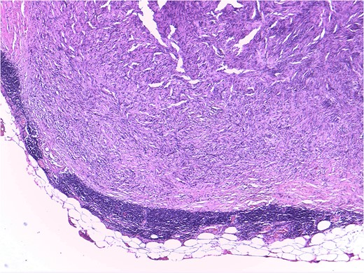
Mesenteric node tumor microscopically constituted by a spindle cell proliferation without atypia, mitosis or necrosis, displacing the normal limphatic tissue toward periphery.
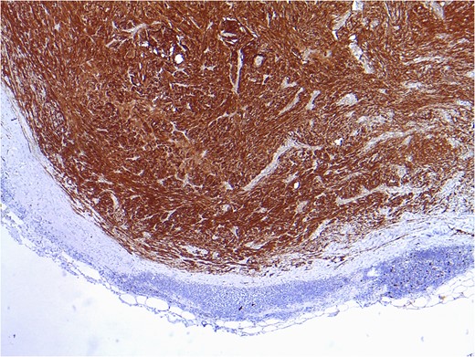
Positive result after immunohistochemical study for vimentin and protein S100, compatible with intranodal schwannoma.
DISCUSSION
Schwannomas are benign neurogenic tumors that originate from Schwann cells. These cells constitute the myelin sheath covering neuronal axons. Lymphatic affectation (within lymph nodes) is extremely rare and poorly described in the literature. The lesions to beware of in the differential diagnosis are: metastatic tumor cells, including spindle cell sarcoma, spindle cell carcinoma and melanomas.
Among the benign lesions, the most important distinction must be made with the miofibroblastoma, whose characteristics include hemorrhage focuses and areas of starry arranged collagen. While miofibroblastomas show a positivity result for actin immunohistochemistry, they do not do it for S100 protein, whose positivity is strongly linked to the schwannomas. In our case, the immunohistochemical consistently oriented in the direction of these latter.
It was reported just one case before by Piana, of mesenteric lymph node affectation by schawnnoma in a piece of colonic resection [1]. Remaining reports describe tumors located in retroperitoneal nodes, thoracic region, groin location or neck mass relative to growth lymph nodes [2–10]. In our case, as in the rest of the consulted case reports, lymph node involvement by schwannoma, could not be identified until the histopathological analysis are completed, either preoperative extension studies or macroscopic study of the surgical specimen, could suspect lymph node involvement by this tumor, because, there are not specific characteristics that could make it suspicious. It is also noteworthy that such lymph node involvement occurred in a different one from the affected by adenocarcinoma metastasis.
These tumors are often discovered as an incidental finding. Intranodal schawnomas are extremely rare and <12 cases have been previously described in the literature. The origin of those remains unknown; therefore, as far as our review is concerned, there are not currently theories that could explain lymphatic localization. On the other hand, we have to notice that this type of diagnosis, although anecdotal by its infrequency, highlights the importance of correct microscopic evaluation of lymphatic tissue, included in the pieces of surgical, beyond their macroscopic appearance, because it has not only diagnostic importance, as in our case, but also implications in staging and subsequent treatment if malignant lesions are present.
