


Abstract
Amputations of a traumatic origin are very frequent in developing countries, in the case of Guatemala these are a result of work accidents very closely related to poor work conditions existing for manual workers, as well as social violence and the lack of security that governs society. The present case shows a patient that suffered a left hand amputation at wrist level. Amputated hand was transported swiftly and in adequate conditions, maintaining cold chain at all times until arrival at Hospital for replantation. After 14 months, patient has evolved satisfactorily and obtained functional result of the hand.
INTRODUCTION
Amputations of a traumatic origin are very frequent in developing countries [1], in the case of Guatemala these are a result of work accidents very closely related to poor work conditions existing for manual workers, as well as social violence and the lack of security that governs society.
Once these amputations occurred, the treatment of them is very likely not viable due to lack of medical staff trained in microsurgery in the main national hospitals, added to the almost non-existence of hospital supplies and lack of understanding of the population in general with regards to first aid treatment of the patient and the adequate conditions for the transport of the amputated member once the accident has occurred, circumstances which complicate the replantation.
CASE REPORT
Patient was assaulted with a large knife (‘machete’), and while the assailant was trying to hurt his face, patient defended himself with his left hand and received the full force of the hit in his arm, cutting of the left hand. Relatives who were with the individual at the time placed the hand in a plastic bag, and the plastic bag in ice, transporting the patient and the hand to Hospital Roosevelt (Fig. 1).
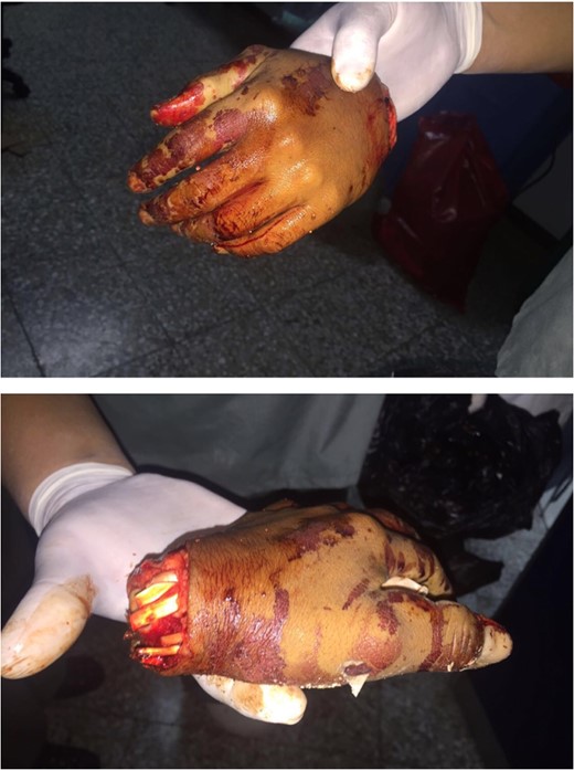
Hand amputation.
Emergency room
Patient was evaluated at entry to the emergency room, conscious and oriented in time, space and person, 15 points in Glasgow scale, vital signs: P/A: 110/70 mmHg, FC: 82 min−1, T: 37.1 °C and 20 breaths per minute, presented a superficial laceration in right side of face, malar region, 8 cm in longitude, transverse, at the level of the left superior extremity; did not present active bleeding of the stump due to blood vessel coagulation, patient is sent to X rays, for AP and lateral projection of the hand and stump (Fig. 2).
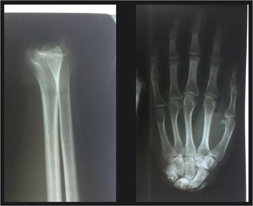
X-rays of the amputated limb
Surgery
Thirty minutes after entry into the ER patient is taken into surgery, where he is given general anesthetic, and subjected to debridement and intense wash of the amputated hand and stump, using surgical soap Gluconate of Clorhexidine and sterile saline solution (10 L).
We proceed to insert K.W. 1.8 mm, running through the distal radius to the carpus (ulnar side) and another from the distal radius to the other side (radial side); once the bone stabilization was obtained, we proceed to identity the ulnar artery and regularize its walls, both of the stump as well as in the hand and we proceed to the anastomosis using nylon 9-0, placing simple separate stitches, clamp is liberated and good perfusion is evidenced; we then proceed to find a vein in the dorsal forearm and in the hand, we dissected the proximal segments and due to a deficit in length we decide to take a vein graft in the dorsal forearm (7 cm in longitude), this is interposed between the ends; we then performed an anastomosis with stitches using nylon 9-0, there was a good flow (venous return) when we released the clamps. We find the ulnar and median nerve stumps and we perform a nerve repair using single stiches with nylon 7-0 along the circumference of both nerves (Fig. 3).
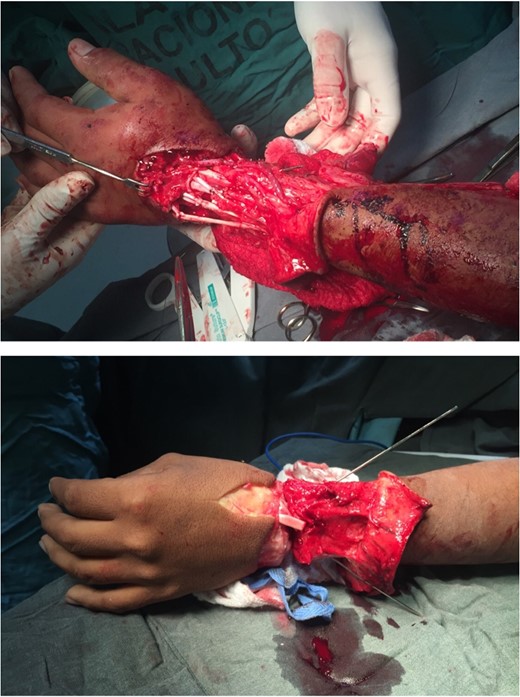
Reimplantation of the hand, with adequate blood supply and reconstruction of tendons.
We then find the flexor and extensor tendons in the forearm and hand and we proceed to repair with Kestler type stitches using nylon 4-0. Skin closure with 4-0, on the radial side of the wrist, palmar side there is a defect of ~4 cm in diameter whereby it is necessary to take and place a full thickness graft for which we use the anterior side of the elbow as donor area.
Monitoring every hour through pulse oximeter. First 48 h all indicators are above 95% in addition evaluation continues on capillary filling (always <2 s in all fingers), uniform temperature and similar coloring to that of the right hand. Evolution of patient was satisfactory, heparin was not used under molecular weight or derivatives, patient did not present immediate post-operative complications and was discharged from hospital 2 weeks after surgery.
Eight months after surgery he was taken into surgery to perform a tenolysis of the flexors. Patient attended physiotherapy at the hospital three times a week in a much disciplined manner for 14 months after surgery.
DISCUSSION
Since 1962, when the first hand replantation was performed in Boston, many cases have been reported and much has been written about limb replantation. Indications in essence continue to be the same and there are no significant variations in concerns [1, 2].
The arrival of new supplies, sutures, microsurgical instruments and high definition microscopes have improved the outcome of these kinds of procedures [2–5].
Despite the high incidence of thrombotic accidents in the first 48 h post replantation (80%), there is not enough evidence to support the use of heparin and derivatives as a rule during the post operatory [4–6]. There are many studies prone to the use and others that concluded that it is not necessary. Nevertheless there are many observations in studies that mention the importance of the surgeon’s technical skills in order to have less thrombosis related complications and better results.
It is important to mention that there are very few reports and information of hand replantation’s in adverse scenarios such as the one presented in this case study (no microscopes available to perform a vascular anastomosis and nerve connections) [7, 8]. Due to the absence of a microscope, the surgery was performed with loupes 4.5; vascular anastomosis was performed with nylon 9-0 due to the lack of microsurgery sutures in the hospital, which was provided by the ophthalmology unit, with which we were able to unite the artery and the veins. During surgery, milliliters of saline solution with two thousand units of heparin were used to irrigate the vascular connections.
In the US in recent years, specialized centers have been created for the treatment of amputations, which nowadays are frequent in number, however, replantation procedures are not a common procedure due to the high cost, prolonged follow up and complications such as failure, infections, and in some cases death [1, 5, 8].
In the early 80s a boom of ectopic replantation began, indicated for cases with extensive damage in stump in soft tissue, contamination and infections, with a treatment consisting of several debridement’s, antibiotics therapy, repair of tissues and connection of the amputated segment into vessels of the abdominal wall, and after recovery, around the 10th or 12th week, standard replantation is performed [9, 10].
Finally it is important to mention studies showing the superiority of the replantation over the use of prosthesis in patients in which replantation was impossible to perform, without of course seeing the future when we talk about the next generation of prosthetic of extremities.
SUPPLEMENTARY DATA
Supplementary material is available at the Journal of Surgical Case Reports online.
CONFLICT OF INTEREST STATEMENT
None declared.
