


Abstract
Rectus sheath hematoma (RSH) is an increasingly common clinical condition in our hospitals due to the increasing use of anticoagulant therapies for various purposes among our patients. Treatment of spontaneous RSH is generally conservative. For continued bleeding, interventional radiologic identification and subsequent embolization is an effective option. Surgery usually involves significant morbidity and is considered a technique of last resort. In this case report, we describe the case of middle aged female who developed abdominal compartment syndrome (ACS) from a large RSH that had extended into the retroperitoneum. The patient underwent abdominal decompression with removal of the hematoma and subsequently fared very well. Patients with large RSHs extending into the retroperitoneum should undergo constant monitoring of their abdominal pressures for early detection and treatment of potentially deadly condition of ACS.
INTRODUCTION
Rectus sheath hematoma (RSH) is an increasingly common problem due to the increasing use of anticoagulation for medical conditions including atrial fibrillation (A-Fib), vascular bypass and mitral valve replacements. RSHs are frequently misdiagnosed or underdiagnosed because of the non-specific clinical presentations and can mimic other causes of abdominal pain. While relatively infrequent, it is usually managed conservatively and has a generally favorable outcome [1]. We report a case of a large RSH extending into the retroperitoneal space resulting in abdominal compartment syndrome (ACS).
CASE REPORT
A 49-year-old female presented to the emergency department with a one-day history of abdominal discomfort. She had a history of factor V Leiden mutation with multiple pulmonary embolisms (PE) and deep venous thrombosis for which she was on long-term warfarin therapy. Her other co-morbidities included hypertension, morbid obesity, atrial fibrillation, remote subdural hemorrhage and mitral valve replacement.
On examination she was pale, had a blood pressure of 97/52 mm Hg and heart rate of 74. On abdominal examination, she was distended and tender in all four quadrants. Her hemoglobin (Hb) was 4.7 g/dl, white blood cell count was 11.4 × 10⁹/l and platelet count was 146 × 103/mcl. Her international normalized ratio (INR) was 1.5, which was reversed effectively with two units of fresh frozen plasma. She was transfused five units of packed cells improving her hemoglobin from 4.7 to 7.8 g/dl. An emergency contrast CT scan of abdomen and pelvis was performed which showed a large right-sided RSH extending into the space of Retzius (Fig. 1).
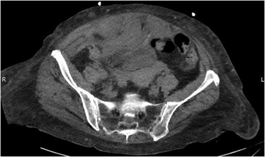
Abdominal CT scan showing right-sided RSH.
The following day the patient’s urine output declined progressively and she subsequently developed acute renal failure. A urinary catheter was placed but significant resistance was encountered secondary to her locally distorted anatomy from the hematoma. A repeat non-contrast CT abdomen and pelvis was therefore obtained which showed further extension of the RSH into the space of Retzius with retroperitoneal extension causing hydronephrosis (Fig. 2).
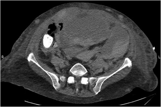
Abdominal CT scan showing the RSH extending into space of Retzius and retroperitoneum.
Her intra-abdominal bladder pressure (IAP) was noted to be 31 mm Hg (Grade IV intra-abdominal hypertension). The patient was emergently taken to the operating room (OR) for abdominal decompression. Necrotic rectus muscle was noted when the fascia was transected. Two liters of organized clot and blood was removed which had dissected into the retroperitoneal space bilaterally. After the damage control laparotomy, the patient was admitted to the surgical ICU with temporary abdominal wall closure achieved by application of Abthera™ ‘Vacuum Assisted Closure’ (KCI, San Antonio, TX, USA). Patient was taken back to the OR within 24 h for re-exploration and abdominal fascial closure. She recovered very well from her surgery with normalized bladder pressures and complete recovery of renal function. A repeat CT scan on postoperative Day 10 showed complete resolution of her rectus sheath and retroperitoneal hematomas (Fig. 3).
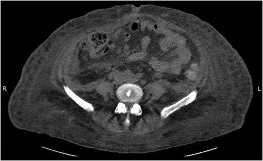
Abdominal CT scan showing complete resolution of rectus sheath and retroperitoneal hematomas.
DISCUSSION
RSH is an increasingly common clinical condition in our hospitals due to the increasing use of anticoagulant therapies for various purposes among our patients. The structural abnormality usually entails a collection of blood principally below the level of the arcuate line. These patients most commonly present with abdominal pain, skin bruising and Fothergill sign—‘an abdominal mass that does not cross the midline and remains palpable when the rectus abdominal muscle is contracted [2]’. Although the exact incidence is unknown [3], Klingler et al. found an incidence of 1.8% among 1257 patients who underwent ultrasonography for acute abdominal pain or unclear acute abdominal disorder [4]. Warfarin, un-fractioned and low-molecular heparin and novel oral anticoagulants (NOACs) have all been implicated in the development of RSH [5].
Treatment of spontaneous RSH is generally conservative which includes analgesia, fluid resuscitation, blood transfusion for symptomatic anemia, correction of the coagulopathy and treatment of the underlying condition. For continued bleeding, interventional radiologic identification and subsequent embolization is an effective option. Surgery usually involves significant morbidity and is considered a technique of last resort.
ACS defined as sustained IAP of 20 mmHg or more associated with new organ dysfunction, is an extremely deadly condition with mortality rates from 29 to 62% [6]. O’Mara et al. in 2003 reported first case of ACS from RSH [7]. Various authors have since then reported ACS secondary to either rectus sheath or retroperitoneal hematoma. Our case if the first of its kind in which ACS results from an RSH hematoma extending into the retroperitoneal space.
An overall review of the literature shows that patients with RSH who develop ACS had poor outcomes if they underwent conservative management.
Jafferbhoy et al. reported case of development of ACS in middle-aged male with RSH secondary to being on Coumadin for aortic valve replacement. He did not undergo surgery as it was felt that this would release the tamponade effect. The patient deteriorated overtime and then expired. Similarly, Kayrak et al. reported that the case of ACS in a 67-year-old female with RSH whose family refused operative intervention. The patient finally succumbed to her disease [8]. In comparison, there was no mortality among the patients who underwent surgery for ACS from RSH or retroperitoneal hematoma [9, 10].
Many of the effects of ACS are clinically indistinguishable from those of other common entities related to critically ill patients, and large hematomas and blood loss—with their abdominal pain, metabolic acidosis, decreased urine output and decreased cardiac output—present similarly to ACS. In these patients, it is probable that the influence of an elevated IAP is not infrequently missed. As a result, clinicians must possess a high index of suspicion and monitor IAP frequently.
CONCLUSION
A large RSH is a severe and potentially fatal complication of anticoagulant therapy. These patients require constant monitoring of hemodynamic and ventilatory parameters since they are at risk for developing ACS. We should have a lower threshold of taking these patients to the OR.
CONFLICT OF INTEREST STATEMENT
None declared.
