


Abstract
Dieulafoy's lesion, a dilated aberrant submucosal vessel which erodes the overlying epithelium, is a relatively rare but potentially fatal cause of gastrointestinal (Gl) bleeding. The esophagus is a very rare location for the lesion. Here we present a case of massive upper GI bleeding, secondary to this remarkably rare occurrence, which was amendable to endoscopic intervention.
Introduction
Dieulafoy's lesion was first described by Gallard in 1884. Originally named the exulceratio simplex, it was named after the French surgeon Georges Dieulafoy 14 years after its initial discovery. Also referred to as ‘cirsoid aneurysm’, the Dieulafoy's lesion consists of a dilated submucosal vessel that causes significant bleeding when it erodes the overlying epithelium, without primary ulcer leading to its exposure. The lesion is most commonly located in the lesser curvature of the stomach, with infrequent occurrences in the small bowel and the colon. The esophagus is an exceedingly rare location for this lesion, with few case reports published in the literature. The pathophysiology of the lesion is believed to be congenital in nature, with rare reports of its occurrence in neonates and young infants. In most published series, however, particular comorbidities appear to have very strong correlation with the Dieulafoy lesion, most notably; cardiovascular disease, hypertension, chronic renal disease, diabetes mellitus and excessive alcohol abuse. Collectively, these comorbidities have been associated with almost 90% of patients having this condition.
Case Report
A 55-year-old male with history of intellectual disability presented to our institutions emergency room after being found by his father to be obtunded and unresponsive. Upon presentation to the patient's room, the surgical team noted that the patient was vomiting large volumes of bright red blood, and was significantly tachycardic to the 140s and hypotensive. Upon questioning, patient's father reported that he is usually communicative, and that he had now been unresponsive for many hours.
Past medical history: diabetes mellitus type II, hypertension, hyperlipidemia, alcohol and polysubstance abuse.
On examination, the conjunctiva appeared pale, and there was minimal capillary refill.
The abdomen was soft, not distended, no rebound; with large amounts of blood noted around the mouth and staining the patients clothes. Following initiation of nasogastric suctioning, saline lavage revealed frank red blood on return.
The patient was emergently resuscitated in the Emergency Department with placement of large bore intravenous access and initiation of the institution's massive transfusion protocol. He was subsequently transferred to the surgical intensive care unit where he continued to have copious amounts of hematamesis. The patient was intubated for airway protection, and emergent esophagogastroduodenoscopy (EGD) was performed at the bedside.
EGD revealed active pulsatile bleeding from an exposed vessel in the distal portion of the esophagus without underlying ulceration (Fig. 1). Attempts were made at epinephrine injection sclerosis; however, bleeding was finally amended with resolution clip placement (Fig. 2).
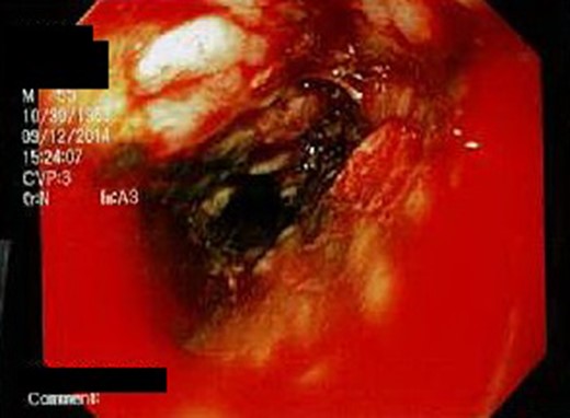
Ectatic vessel can be seen at the 3 o'clock position.
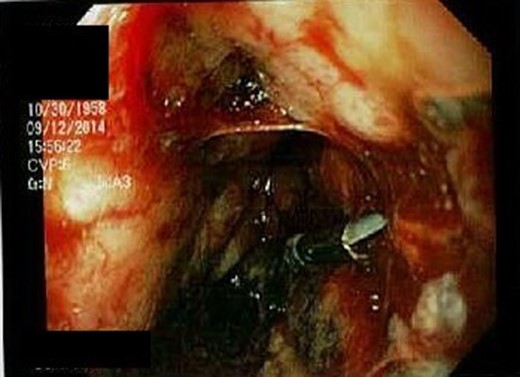
Resolution of hemorrhage following placement of hemostatic clip.
Following establishment of hemostasis, the patient was maintained on PPI drip in the SICU and progressively stabilized. He was extubated and eventually discharged home in stable condition.
Discussion
One of the more rare causes of upper gastrointestinal (GI) bleeding is Dieulafoy's lesion, which is responsible for ~1.5% of acute upper GI bleeding [1]. Paul Georges Dieulafoy named the lesion after he described a series of 10 patients who presented with massive hematemesis due to a bleeding gastric vessel without associated ulceration in 1897. While initially only described in the stomach, Dieulafoy lesions have presented at several other anatomic locations. Of note, 75–90% occur in the proximal stomach, though small bowel, colon, ano-rectal and extra-intestinal lesions have also been described [2].
A Dieulafoy's lesion is a congenital arteriovenous malformation characterized by an ectatic submucosal artery; if eroded it may lead to pulsatile bleeding. Anatomically, arteries should narrow as they approach the mucosa, but a Dieulafoy artery maintains a persistent caliber through the submucosa, leading to the eponym of ‘caliber-persistent artery’ [3]. The mechanisms causing the tortuosity and the persistence of the large-sized submucosal arteries remain unknown, though bleeding is thought to be secondary to erosion caused by the pulsations of the artery and not by a primary ulceration of the mucosa [2].
Endoscopy has become the mainstay of diagnosis and treatment for Dieulafoy lesions. The lesion appears as a stream of arterial blood emanating from what appears to be grossly normal mucosa, though a minute defect should be present. Up to 71% of initial endoscopies are diagnostic only if there is active hemorrhage of at least 0.5 mL/min [4]. However, in 6% of patients, the intermittent nature of the bleeding often requires multiple endoscopies to establish a definitive diagnosis like in our patient. Due to the subtlety of the lesion, endoscopic criteria have been established for diagnosis of a Dieulafoy lesion; endoscopic visual criteria that must be met include (i) active arterial spurting or oozing from a small (<3 mm) defect in the mucosa, (ii) visualization of a vessel protruding from a slight defect or normal mucosa and/or (iii) a fresh blood clot adherent to a defect of normal mucosa as described by Dieulafoy [5].
With the advent of endoscopy, multiple modalities may be used for treatment and since 1986 this has become the mainstay for treating these lesions. The endoscopist may choose to band the lesion, use an endoscopic clip, as we had in this patient, use thermocoagulation or inject epinephrine into the lesion. If endoscopy fails to diagnose the lesion, angiography may be employed and the lesion may be embolized. If minimally invasive approaches fail, surgical intervention may be warranted if the patient continues in extremis. Surgery is reserved as a last resort when the location of the lesion is known and previously attempted therapies have failed. Intraoperatively, the lesion may be amendable to oversewing or a wide local excision may be performed, which is preferable given that simple oversewing has shown to be associated with a greater incidence of recurrent bleeding [6].
Conflict of interest statement
None declared.
