Abstract

It might be possible to remove thymic tumors with minimal invasion of the left brachiocephalic vein (BCV) using an advanced videoscopic technique; simple resection of this vessel can be achieved via such an approach. However, tumor invasion of the superior vena cava or right  BCV requires angioplasty or reconstruction, both of which are difficult to perform in videoscopic procedures. We report a case of invasive thymoma with localized invasion of the great vessels at the junction of the left BCV and superior vena cava. An L-shaped mini-sternotomy combined with a videoscopic approach allowed thymectomy with safe vesselplasty of the involved vessels.

Introduction

We report a case of invasive thymoma with localized invasion of great vessels at the junction of the left brachiocephalic vein (BCV) and superior vena cava. An L-shaped mini-sternotomy combined with video-assisted thoracic surgery (VATS) allowed thymectomy with safe vesselplasty of the left innominate vein.

Case
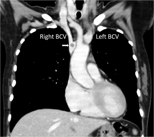
A 52-year-old female was referred to our hospital diagnosed as myasthenia gravis (MG). She had a right-sided anterior mediastinal tumor suspected of thymoma (diameter, 34 mm) with a polyp-like intravascular extension into the left BCV (Fig. 1). Preoperatively, she underwent myasthenic crisis twice and required tracheostomy and mechanical ventilation for several weeks. Prior to that, she had been receiving prednisolone 40 mg daily, and continued high-dose steroid therapy for several weeks after control of the myasthenic crises.
Chest computed tomography showing a nodule at the junction of the right and left BCV (arrow).
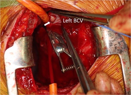
Figure 1:

Chest computed tomography showing a nodule at the junction of the right and left BCV (arrow).

To remove her tumor, right-sided VATS and L-shaped mini-sternotomy were performed with the patient in a right hemi-lateral supine position. Three 5-mm ports and one 11.5-mm port were placed in the right chest for VATS. Under video view, the intrathoracic field was assessed after which the thymus and anterior mediastinal fat tissues were mobilized from the pericardium along the phrenic nerve and opposite side of the mediastinal pleura, beginning from the diaphragm and going cranially. After this videoscopic procedure, the patient was moved from a hemi-lateral to supine position. The sternum was then divided in the midline from the sternal notch to the second intercostal space and its right half transected horizontally, creating an L-shaped sternal incision. After placement of retractors, the remaining upper part of the thymus was manipulated to expose both BCVs and the superior vena cava (SVC), and tourniquets and side clamps were applied to these vessels. The junction of the right and left BCV was then transected and thymectomy, including the intravascular component and involved vessel wall, achieved (Fig. 2). Negative surgical margins were confirmed by intraoperative pathological examination, after which the vessel walls were repaired directly without an artificial graft through the mini-sternotomy. At the end of the procedure, a 20-Fr chest tube was placed in the thoracic cavity and the wounds were closed. Pathological finding was type B1 thymoma [1]. The chest tube was removed on postoperative day 1. No complications occurred. The patient was then treated with radiation therapy (52 Gy). There was no recurrence 64 months after surgery.
Intraoperative photo showing the tumor extending into the left BCV (arrow).
Figure 2:

Intraoperative photo showing the tumor extending into the left BCV (arrow).

Discussion

It is commonly believed that only non-invasive thymomas within 5 cm in diameter can be managed by VATS. However, recent advances in videoscopic techniques have allowed resection of localized thymomas invading the lungs, phrenic nerve and pericardium [2]. Because simple resection of the left BCV is possible, VATS can be performed in cases with minimal invasion of this vessel. On the other hand, when tumor invasion has occurred at the level of the right BCV or SVC, angioplasty or vessel construction is required. These procedures are difficult to perform in videoscopic procedures but can be achieved by the addition of minimally invasive surgery, thus minimizing the incidence of sternal dehiscence and infections, which occur in 0.5–5% of open heart surgeries [3]. The risk of sternal complications was likely greater in the present case because she was receiving high-dose steroid therapy. Sternal instability would have caused serious respiratory complications in this patient, who may again have developed an MG crisis. We, therefore, aimed to minimize surgical damage to the thorax. A combined mini-sternotomy/VATS technique for performing thymectomies in patients with MG and minimally invasive treatment of cardiovascular disease has been reported [4, 5]. In our case, an L-shaped sternotomy provided sufficient surgical exposure of major veins and allowed accurate angioplasty of these vessels. If the surgical field is inadequate for this procedure, then conversion to a reversed T or full sternotomy is easily achieved [5]. Adding mini-sternotomy to VATS can facilitate angioplastic procedures on major vessels.

Conflict of interest statement

None declared.

References

1

World Health Organization Classification of Tumours
.
Pathology and Genetics of Tumour of the Lung, Pleura, Thymus and Heart
.
Lyon
:
International Agency for Research on Cancer (IARC) Press
,
2004
.

2

Pennathur
A
,
Qureshi
I
,
Schuchert
MJ
,
Matthew
JS
,
Dhupar
R
,
Ferson
PF
, et al. .
Comparison of surgical techniques for early-stage thymoma: feasibility of minimally invasive thymectomy and comparison with open resection
.
J Thorac Cardiovasc Surg
2011
;
141
:
649
701
.

3

Olbrecht
VA
,
Barreiro
CJ
,
Bonde
PN
,
Williams
JA
,
Baumgartner
WA
,
Gott
VL
, et al. .
Clinical outcomes of noninfectious sternal dehiscence after median sternotomy
.
Ann Thorac Surg
2006
;
82
:
902
7
.

4

Grandjesn
JG
,
Lucchi
M
,
Mariani
MA
.
Reversed-T upper mini-sternotomy for extended thymectomy in myasthenic patients
.
Ann Thorac Surg
2000
;
70
:
1423
5
.

5

Autschbach
R
,
Walther
T
,
Falk
V
,
Diegeler
A
,
Metz
S
,
Mohr
FW
.
S-shaped in comparison to L-shaped partial sternotomy for less invasive aortic valve replacement
.
Eur J Cardiothoracic Surg
1998
;
14
:
S117
21
.

This is an Open Access article distributed under the terms of the Creative Commons Attribution Non-Commercial License (http://creativecommons.org/licenses/by-nc/4.0/), which permits non-commercial re-use, distribution, and reproduction in any medium, provided the original work is properly cited. For commercial re-use, please contact journals.permissions@oup.com