Abstract

Primary cardiac tumour is a rare entity as secondaries in the heart are more common. A 2-year-old child was having repeated respiratory tract infection with poor oral intake and poor activity for 3 months. His symptoms progressed from New York Heart Association (NYHA) Class II to IV. On evaluation he had an intracardiac mass with extracardiac extension. Emergency tumour excision under deep hypothermic circulatory arrest was performed with provisional diagnosis of sarcoma. But Serum markers, histopathological examination and immunohistochemistry confirmed diagnosis of yolk sac tumour. Postoperative recovery was uneventful and the child was receiving adjuvant chemotherapy. Extensive literature review revealed only four cases of primary intracardiac yolk sac tumour published till date. Our case report is unique, in that intracardiac tumour had extracardiac extension by infiltration through right atrial wall. Previous four reports mention purely intracardiac mass.

Introduction

Primary cardiac tumours are a rare entity whose incidence according to surgery and autopsy reports is 0.3–0.7% of all cardiac tumours [1]. Germ cell tumours are benign or malignant neoplasms arising from primordial germ cells which can be gonadal or extragonadal in origin. Primary intracardiac yolk sac tumour is extremely rare, only four cases being reported till date. We report probably fifth case.

Case Report

A 2-year-old male child was brought to hospital with respiratory distress since last 2 weeks. He was having repeated respiratory tract infection since last 3 months along with poor oral intake and poor activity. His symptoms progressed from New York Heart Association (NYHA) Functional Class II to IV over last 2 weeks. On investigation at another hospital he was diagnosed with intracardiac mass having extracardiac extension. Parents brought the child to our hospital for further management.
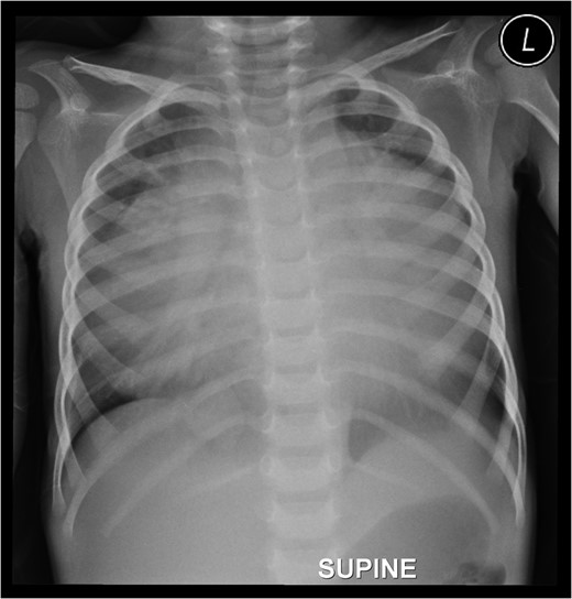
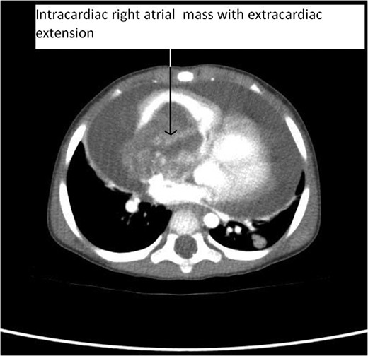
On clinical examination patient was in severe respiratory distress with respiratory rate of 48/min. He was pale with facial puffiness and periorbital oedema. Pulse rate was 136/min, regular having low volume. Jugular venous pressure was 7 cm. Breath sounds were normal. Abdomen was distended nontender with 5 cm hepatomegaly. Chest roentgenogram revealed huge cardiomegaly with inhomogeneous margins suggestive of massive pericardial effusion, cardiothoracic ratio of 0.8 with clear lung fields (Fig. 1). Electrocardiography showed low voltage sinus tachycardia with rate of 136/min and electrical alternans. Echocardiography revealed massive pericardial effusion with an intracardiac mass occupying whole right atrium (RA) and obstructing tricuspid valve with ball valve mechanism (Fig. 2, Video 1). Computed tomography (CT) of thorax showed enlarged RA almost totally occupied by huge solid mass of heterogeneous enhancement and lobulated outlines measuring 7×7×6 cm and extending through tricuspid valve. Superolaterally mass is infiltrating superior vena cava (SVC)–right atrium (RA) junction and extending up to bracheocephalic vein (Fig. 3). It also showed massive pericardial effusion. Right lung in lower lobe had two solid nodules the largest measuring about 1.7×1.7 cm. Provisional diagnosis of sarcoma was made as it is the most common malignant cardiac tumour in children. Emergency surgery was undertaken considering NYHA Class IV symptoms and worsening clinical status.
Preoperative chest roentgenogram showing cardiomegaly and massive pericardial effusion.
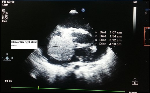
Figure 1:

Preoperative chest roentgenogram showing cardiomegaly and massive pericardial effusion.
Preoperative 2DECHO showing right atrial mass.
Figure 2:

Preoperative 2DECHO showing right atrial mass.

CT Thorax showing intracardiac right atrial mass with extracardiac extension.
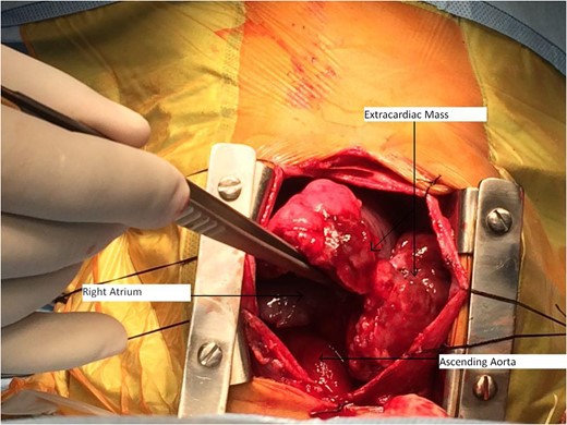
Figure 3:

CT Thorax showing intracardiac right atrial mass with extracardiac extension.
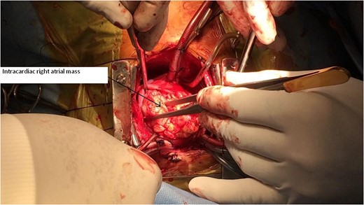
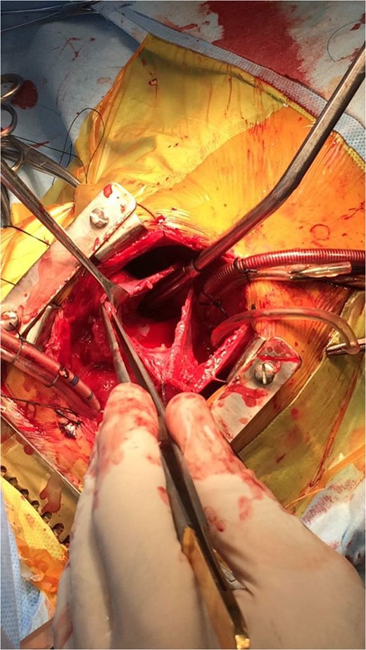
Median sternotomy approach was used. Thymus was normal. Pericardium was stretched and bulging out. Haemorrhagic effusion of 150 ml was drained. Mass of size 7×7×4 cm was overlying RA and enveloping Aorta and Pulmonary arteries (Fig. 4). Mass was free from pericardium and was pushing the RA downwards and both ventricles to left side. Extracardiac mass excised in piecemeal to make sufficient space for aorta and SVC cannulation. Mass was attached to RA. High SVC cannulation done as mass was invading SVC lumen. Cardiopulmonary bypass established. Deep hypothermic circulatory arrest achieved along with cardioplegic arrest; RA opened. Intracardiac mass was almost fully occupying the RA and was extending to right ventricle through tricuspid valve (Fig. 5, Video 2). It was attached to the SVC–RA junction and trabecular portion of RA. Resection of intracardiac tumour done except at its attachment to SVC–RA junction where it had infiltrated wall (Fig. 6). Tricuspid valve was fairly competent. Postoperatively he had smooth recovery with symptomatic improvement from NYHA Class IV to Class I.
Intraoperative image of extracardiac mass.
Figure 4:

Intraoperative image of extracardiac mass.

Intraoperative image of intracardiac right atrial mass.
Figure 5:

Intraoperative image of intracardiac right atrial mass.

Right atrium after resection of intracardiac mass.
Figure 6:

Right atrium after resection of intracardiac mass.

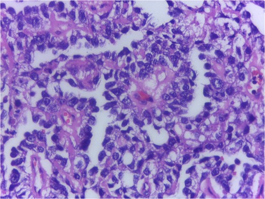
Grossly tumour was greyish, vaguely nodular having soft to firm consistency. Cut section was greywhite and glistening with focal yellowish and haemorrhagic areas. Specimen was extensively sampled for histopathological examination. Microscopic examination revealed neoplastic tissue composed of pleomorphic cells with ovoid to irregular vesicular to hyperchromatic nuclei, variably prominent nucleoli and scanty to moderate amount of cytoplasm in microcystic and glandular pattern predominantly along with nests and trabeculae. Several Schiller Duval bodies were seen (Fig. 7). High nucleocytoplasmic ratio noted. Necrosis haemorrhage and dispersed foci of calcification seen. Scattered mitotic figures were seen (10–12/hpf). Periodic acid–Schiff (PAS) stain highlighted the hyaline globules.
Histology H&E 400× showing Schiller Duval bodies.
Figure 7:

Histology H&E 400× showing Schiller Duval bodies.

Serum markers on day of surgery were Alpha Feto Protein (AFP) 131 146 ng/ml (normal range 0–20 ng/ml), Beta subunit of Human Chorionic Gonadotropin (beta hCG) 0.5 mIU/ml (normal range < 2 mIU/ml), Lactate Dehydrogenase (LDH) 1231 U/L (normal range 0–850 U/L)
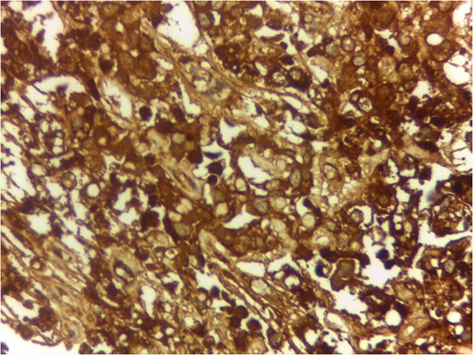
Immunohistochemistry (IHC) examination revealed neoplastic cells were positive for AFP (Fig. 8), Glypican 3 CK and negative for CD30, beta hCG, D240, Epithelial Membrane Antigen.
IHC for Alpha Feto Protein 400×.
Figure 8:

IHC for Alpha Feto Protein 400×.

Serum markers, histopathological examination and IHC all three confirmed diagnosis of yolk sac tumour.

Postoperatively Serum AFP was elevated to 830 900 ng/ml. Early chemotherapy was started with Bleomycin, Etoposide and Cisplatin (BEP) regimen. The child was tolerating chemotherapy well. After second cycle of chemotherapy Serum AFP level dipped to 2025 ng/ml showing good response to chemotherapy. Ultrasonography of abdomen pelvis and testis was normal prior to chemotherapy. We will review patient after each chemotherapy cycle and in long term for prognosis and recurrence if any.

Discussion

Primary cardiac tumour is rare. Only 25% of primary cardiac tumours are malignant. Survival rate for malignant primary cardiac tumours without surgical resection at 9–12 months is only 10%. Sarcomas constitute 75% of malignant primary cardiac tumour. Germ cell tumour is rare. Germ cell tumours are due to abnormal differentiation of foetal germ cells that arise from the foetal yolk sac. Normal migration of these germ cells may cause gonadal tumours, i.e. ovary and testis, whereas abnormal migration produces extragonadal tumours. Most cardiac germ cell tumours are teratomas. Yolk sac tumour is extremely rare. Surgical resection followed by chemotherapy is preferred treatment for yolk sac tumour. Only four cases of primary intracardiac yolk sac tumour are published till date in medical literature. Those are listed in Refs [25]. Three other cases of yolk sac tumour are reported but they were extracardiac being in pericardial cavity [6]. We are probably reporting fifth case of primary intracardiac yolk sac tumour.

Our case report is unique, in that intracardiac tumour had extracardiac extension by infiltration through right atrial wall. Previous four reports mention purely intracardiac mass. The rarity in the literature of such pathological occurrence makes our case report very unique.

In conclusion, as far as the rarity of the tumour is considered, measures such as thorough preoperative evaluation and preparation, intraoperative decision making and surgical expertise, proper postoperative chemotherapy are must for better outcome.

Acknowledgements

The authors would like to acknowledge Dr Ami Maria Emmanuel, MBBS, MD, Sr. Registrar—Pathology Lakeshore Hospital and Research Centre, Kochi, Kerala, India.

Supplementary Material

Supplementary material is available at the Journal of Surgical Case Reports online.

Conflict of Interest Statement

None declared.

References

1

Leja
MJ
,
Shah
DJ
,
Reardon
MJ
.
Primary cardiac tumors
.
Tex Heart Inst J
2011
;
38
:
261
2
.

2

Amalaseelan
JV
,
Pieris
DC
,
Munasinghe
M
.
A case of intra cardiac yolk sac tumour
.
Ceylon Med J
2010
;
55
:
53
4
.

3

Melinda
JM
,
Richard
AH
,
William
SF
,
James
WZ.
Intracardiac yolk sac tumor and dysrhythmia as an etiology of pediatric syncope
.
Pediatrics
2004
;
113
:
374
6
.

4

Graf
M
,
Blaeker
H
,
Schnabel
P
,
Serpi
M
,
Ulmer
HE
,
Otto
HF
.
Intracardiac yolk sac tumor in an infant girl
.
Pathol Res Pract
1999
;
195
:
193
7
.

5

Parvathy
U
,
Balakrishnan
KR
,
Ranjit
MS
,
Kuruvilla
S
,
Rao
KR
.
Primary intracardiac yolk sac tumor
.
Pediatr Cardiol
1998
;
19
:
495
7
.

6

Liang
TC
,
Lu
MY
,
Chen
SJ
,
Lu
FL
,
Lin
KH
.
Cardiac tamponade caused by intrapericardial yolk sac tumor in a boy
.
J Formos Med Assoc
2002
;
101
:
355
8
.

This is an Open Access article distributed under the terms of the Creative Commons Attribution Non-Commercial License (http://creativecommons.org/licenses/by-nc/4.0/), which permits non-commercial re-use, distribution, and reproduction in any medium, provided the original work is properly cited. For commercial re-use, please contact journals.permissions@oup.com