-
PDF
- Split View
-
Views
-
Cite
Cite
Thomas Bochmann, Richard Forrester, Jon Smith, Case report: imaging the clinical course of FOPE—a cause of adolescent knee pain, Journal of Surgical Case Reports, Volume 2016, Issue 11, November 2016, rjw178, https://doi.org/10.1093/jscr/rjw178
Close - Share Icon Share
Abstract
Focal periphyseal oedema (FOPE) is a rare MRI finding associated with pain in adolescent patients with very few reported cases. We present a case of FOPE in a 13-year-old girl and the only follow-up imaging available for an isolated presentation of this condition.This article describes a clinical course that correlates well with the imaging obtained. This article describes a clinical course that correlates well with the imaging obtained.
Introduction
We present the case of a 13-year-old girl with left knee pain, an MRI diagnosed focal periphyseal oedema (FOPE) lesion and no other identified injury or morphological abnormality. According to our literature search, this is the only case report that provides follow-up images for a case of isolated symptomatic FOPE.
FOPE is a term used to describe the MRI finding of bone marrow oedema situated around a physis at the time of its fusion, and affects lower limb bones of the adolescent skeleton in either sex [1]. This is a rare finding of uncertain aetiology and significance that has shown itself to be a self-limiting condition [1, 2].
FOPE lesions have so far only been described in patients with pain in the knee. Most often they are active adolescents, and it may occur with or without a definitive history of trauma and associated injury [1, 2].
Proposed theories for the aetiology and significance of FOPE lesions were made when it was initially named in 2011 [1]. However, the volume of evidence currently in existence and the lack of basic science mean that at present these are untested.
A literature search using Pubmed and Medline produced just two papers [1, 2] presenting a total of 15 cases, of which only five had isolated findings of FOPE. The remaining cases all had evidence of concomitant injury, morphological abnormality or musculoskeletal disease complicating the clinical picture.
None of the five cases of isolated FOPE underwent MRI imaging follow-up and so it is difficult to see if their symptoms correlated with changes to the FOPE lesion.
Case Report
The images presented here are those of a fit and active 13-year-old girl with no significant medical history. She complained of intermittent pain, swelling and locking in the left knee of nine months duration.

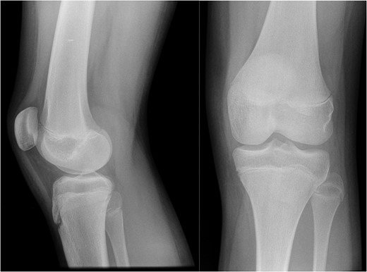
AP and lateral radiographs of the knee from the time of initial injury.
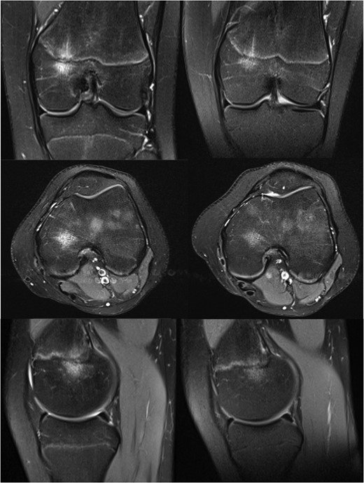
On examination at nine months she had medial tenderness and a positive McMurray's test, raising the suspicion of meniscal injury, for which the initial MRI investigation was performed. This scan showed a focal region of oedema centred in the medial aspect of the distal femoral physis with no other internal derangement, and she was managed with observation.

MRI images of the knee at 9 (left) and 13 months (right) after initial injury.
Her symptoms gradually improved without treatment, and with the significant improvement seen on her MRI scan she was eventually discharged from our care fourteen months after her initial injury.
Discussion
Whilst coining the term FOPE, Zbojniewicz and Laor [1] also attempted to theorize its underlying aetiology. They suggest the possibility of micro-fractures from athletic activity or trauma before going on to conclude that it may be an incidental finding relating to endochondral ossification, which may or may not be painful.
Their final conclusion was based on the preponderance for FOPE lesions to occur in adolescents during physeal fusion coupled with the presence of additional pathology in the majority of cases. However, the apparent correlation with athletic activity or trauma, and the absence of reported asymptomatic cases does not support this conclusion.
The case described here shows a progression of the FOPE lesion towards resolution that is well correlated with the clinical development experienced by our patient. The story clearly relates to trauma in an active adolescent, and is very similar to that described previously.
This suggests that in this case FOPE was the cause of pain and that our findings support the theory that trauma may play a role its aetiology.
This case is in keeping with the previously described cohort of affected patients and supports the current data suggesting that FOPE is a self-limiting condition that does not require any invasive investigation or management.
It provides added insight into the progression of FOPE that has previously not been reported and suggests that trauma is implicit in the development of FOPE lesions.
Conflict of Interest Statement
None declared.


