


Abstract
Perforated peptic ulcer disease remains a relatively frequent emergency surgery presentation. Persistent leak is the most common indication for return to theatre. We present a technique to manage patients in whom a more substantial resection is not possible. A 45-year-old woman underwent initial laparoscopic primary closure of a non-malignant perforated gastric ulcer. This subsequently leaked on return to the UK and had a further graham patch formed via a laparotomy. Unfortunately, the patch repair leaked and at reoperation a wedge excision or distal gastrectomy was not possible given the friability of the tissues and instability of the patient, a transgastric drain and perigastric drain were therefore placed. This created a controlled fistula, which was managed eventually as an outpatient. Transgastric drains in the context of the persistent perforated gastric ulcer leak are a safe way to manage the unstable patient with poor tissues where more substantial surgeries such as a distal gastrectomy are not possible.
INTRODUCTION
Perforated gastroduodenal ulcer remains a common indication for emergency surgery [1].
Perforated peptic ulcer (PPU) is a common surgical emergency that carries high mortality and morbidity rates. Globally, one-quarter of a million people die from peptic ulcer disease each year [2].
In a Danish study of 726 patients treated surgically for PPU between 2011 and 2013, 238 (32.8%) were treated laparoscopically and 178 (24.5%) had a laparoscopic procedure converted to laparotomy. Overall, 124 (17.1%) of the 726 patients underwent reoperation. A persistent leak was the most frequent cause [3].
Regardless of the operative strategy used to manage the initial perforation, a subsequent leak can be a difficult dilemma to manage. As far as the authors are aware, there is no open discussion in the literature on how to manage these difficult patients. The definitive solution is a distal gastrectomy, but this procedure carries significant morbidity and in the unstable patient with poor tissue quality can be disastrous. Indeed, in some cases, it may not be possible to mobilize the duodenum to enable distal gastrectomy to be performed due to the inflammatory response from prolonged exposure to gastric content spillage.
We present an interesting case on how to safely manage this group of patients.
CASE REPORT
A 45-year-old female presented to hospital with abdominal pain of 1-week duration (Day 7) following perforation and peritonitis secondary to a perforated prepyloric gastric ulcer on Day 1 on the antrum of the stomach whilst on holiday in the European Union.
This initial operation had been performed with a laparoscopic repair and primary closure of the ulcer. There was initially an uneventful postoperative period, and the patient returned home.
The patient returned to the UK progressively feeling more unwell with abdominal pain and associated fever. The patient immediately after landing in the UK presented to our district general hospital.
Following review, the patient was started on meropenem with abdominal examination finding peritonism. Imaging demonstrated evidence of free air under the right hemidiaphragm, and the patient was subsequently taken to theatre.
At laparotomy (Day 7), there was two quadrant peritonitis with purulent free fluid. A 2–3-cm pyloric ulcer was identified with loose laparoscopic stitches and free gastric contents leaking from it. The ulcer was found to be partially walled off by the liver and the falciform ligament. The ulcer was closed with vicryl sutures, and omental patch was placed on top via an open upper laparotomy. Biopsies returned showing no malignancy.
Following this second operation, the patient was discharged to intensive care unit (ITU); she initially did well and then was discharged to the ward. She failed to progress on the ward, developing a progressively worsening systemic inflammatory response, and a repeat computed tomography (CT) was undertaken (Day 13). This CT demonstrated a large loculated collection containing fluid and gas anterior to the left lobe of the liver that may be amenable to percutaneous drainage with free fluid throughout the abdomen with upper abdominal free gas. It also demonstrated diffuse fatty infiltration of the liver but no liver abscess. Diffuse fatty infiltration of the liver, but no liver abscess. Following this report, interventional radiological drainage was attempted but failed. Following this, her abdomen was found to be distended and tender with evidence of leak from gastric ulcer perforation.
The decision to take the patient back to theatre was made on Day 16 for an emergency laparotomy for attempted partial gastrectomy and formation of a controlled fistula. The wound was found to have broken down at the level of the fascia with bowel prolapsing out with pus and gastric contents leaking through with the large abscess collection in Left Upper Quadrant and contaminated gastric contents throughout the upper abdomen. The duodenum was unable to be mobilized due to dense peritoneal inflammation and contamination. It was decided to pass a transgastric drain through the abdominal wall and into the non-healing defect in the distal stomach to produce a controlled fistula. A further drain was placed external to the stomach to protect against a leak (Figs 1 and 2).
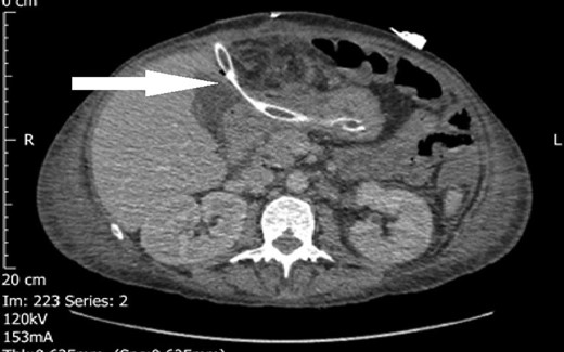
Transgastric drain (marked by arrow).
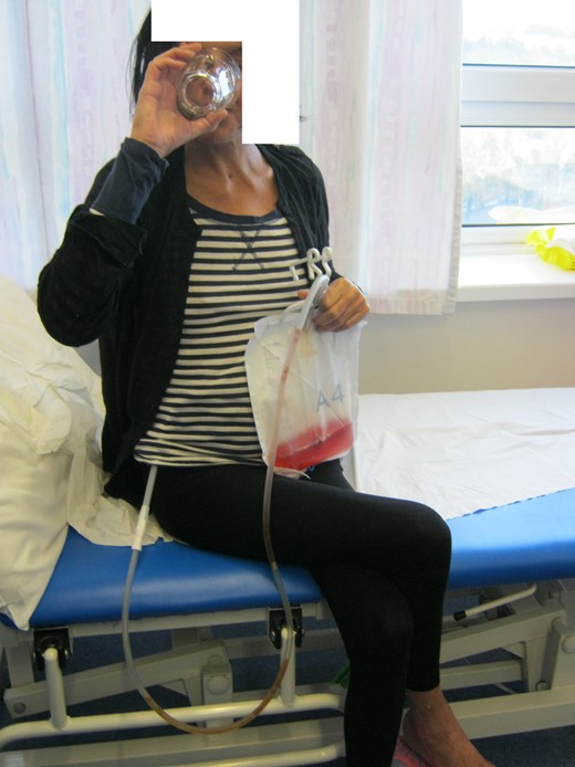
Patient drinking blackcurrant cordial in clinic with passage into her transgastric drain.
Following this, the patient was discharged to ITU. The patient was started on Total Parental Nutrition prior to jejunal feeding. Once the patient was back on the ward, a repeat CT was performed due to raised temperature, which showed no significant collection and likely ascites. The perigastric drain was removed (Day 35) after being cut and having minimal output.
The patient did well following this and was eventually discharged to be followed up in clinic. The gastric bypass drain was removed (Day 57).
She has had a subsequent Oesophago Gastric Duodenoscopy, which was normal and has been discharged from follow-up.
DISCUSSION
The success of medical therapy in the management of peptic ulcer disease now means that the surgery has a very limited role with elective peptic ulcer surgery being virtually abandoned. Emergency presentations of PPU nearly always require surgery. However, in a patient with a small perforation, without peritoneal signs, one can assume the perforation has sealed off physiologically and therefore can be managed conservatively; conversely, pneumoperitoneum and peritonism are absolute indications for surgery [4].
Delaying the initiation of surgery >12 h after presentation is associated with poor outcomes; therefore, when indicated, laparotomy should be performed as soon as possible [5, 6].
Perforated peptic ulcer in contrast to duodenal ulcers, which have a very low cancer risk, will be malignant in 6–14% of cases [7–10]. This affects the decision to patch or resect, however, with the ability to cure gastric lymphoma without resection; many upper Gastro Intestinal surgeons now advocate an initial patch approach with biopsies and subsequent delayed management. Some will still advocate a wedge excision or even distal gastrectomy at the index procedure if the ulcer is in an irregular position or too large with many authors agreeing >1 cm but with no definite measurement at which a graham patch is unlikely to succeed.
The ideal solution for a persistent leak would be wedge excision or distal gastrectomy; however, as is highlighted in the case above, if contamination has been present for some time, this can often present more problems and risk to the patient with friable tissue in the unstable patient. Transgastric and perigastric drain management forming a controlled fistula is a safe way to manage the persistent leak in the non-malignant PPU.
AUTHORS’ CONTRIBUTION
All persons designated as authors have participated sufficiently in the substantial contributions to conception and design or analysis and interpretation of data, drafting of the manuscript or revising it for important intellectual content and final approval of the version to be published.
CONFLICT OF INTEREST STATEMENT
None declared.
ACKNOWLEDGEMENTS
The authors acknowledge and thank all members of the upper gastrointestinal multidisciplinary team including the specialist nurse practitioners, anaesthetists and theatre staff.
