-
PDF
- Split View
-
Views
-
Cite
Cite
James R.L. Davies, Gavin Smith, Andrew J. Cornaby, Teresa Thomas, Michael J. Lamparelli, Delayed recurrence of renal cell carcinoma presenting as a haemorrhoid, Journal of Surgical Case Reports, Volume 2015, Issue 3, March 2015, rjv022, https://doi.org/10.1093/jscr/rjv022
Close - Share Icon Share
Abstract
Metastatic non-colorectal cancer of the anal canal is a rare entity. To date, only four cases have been described in the literature. We present a 76-year-old man who was referred with an unusual perianal lesion. He had a history of renal cell carcinoma 7 years previously. Histologically, the lesion revealed clear cell carcinoma in keeping with metastasis. To our knowledge, this is only the second time a renal carcinoma metastasis to the anal canal has been identified.
INTRODUCTION
Malignancy involving the kidney was estimated to be the 14th most common malignancy worldwide in 2008, with an estimated 273 500 new cases diagnosed [1]. Rates have been found to be higher in more socio-economically developed countries, with an increased risk linked to the male population, smoking and obesity. With this, the incidence of renal cancer is increasing and it is anticipated that in the year 2030 there will be approximately 466 000 new cases diagnosed [1].
About 85% of malignancy involves the renal parenchyma, with the remaining 15% affecting the urothelium. Of the malignancies involving the parenchyma renal cell carcinoma (RCC) is the most common, with clear cell carcinoma as the most common subtype.
CASE REPORT
We present a 76-year-old man who was referred to colorectal clinic with an unusual perianal lesion. A past medical history included sarcoidosis, atrial fibrillation, hypertension, chronic kidney disease and clear cell carcinoma of the right kidney, for which he underwent right-sided nephroureterectomy 7 years previously.
Histology at that time revealed a 34-mm diameter RCC with a 10-mm satellite focus in the adjacent cortex. There was invasion of the renal vein with extension into the perinephric tissue, and Furhman grade was 4.
The patient received a regular urological follow-up and surveillance imaging. In 2011, a CT thorax noted lung nodules suspicious for metastatic disease that were kept under surveillance. In 2013, it was noted that there was a soft tissue mass in the small bowel mesentry.
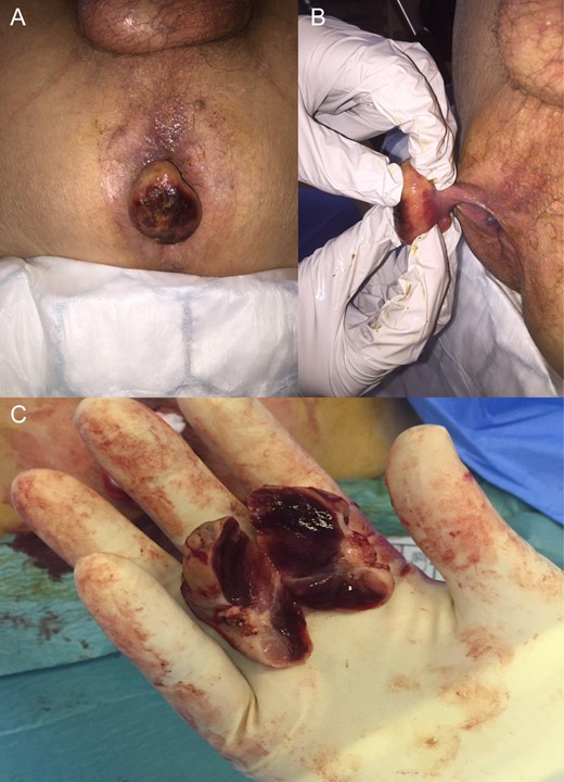
The perianal lesion was removed under general anaesthetic. At time of surgery, the lesion was clearly not a haemorrhoid, although there were grade III haemorrhoids present, and on its cut surface had the appearance of renal tissue. The pedunculated appearance (B) and cut surface (C) are evident in Fig. 1.

Photographs were taken by M.J.L. with permission. (A) Mass evident in lithotomy position. (B) Stalk at base of lesion. (C) Cut surface revealing renal-type tissue.
On histological examination, the sections revealed a covering of hyperplastic squamous epithelium which was focally ulcerated. The core of the nodule consisted of epithelial tumour cells with moderately hyperchromatic nuclei and abundant clear cytoplasm, forming nests and tubules with intervening congested thin-walled vessels.
On immunohistochemistry, testing the tumour cells was positive for CK8/18, EMA, CD10 and vimentin, and negative for CK7. These findings were in keeping with the diagnosis of metastatic RCC of conventional clear cell type.
Given the above findings, the patient underwent full imaging again. Unfortunately, CT head and subsequent MRI revealed a 1.2-cm lesion adjacent to the occipital horn of the left lateral ventricle, with surrounding oedema, suggestive of metastasis. He continues to receive ongoing multidisciplinary team follow-up and management.
DISCUSSION
The most common sites for RCC metastases/recurrence are lung, bone, liver, brain and in the renal fossa; however, there are many reports in the literature of metastasis to other organs including gallbladder, pancreas, gastrointestinal tract, thyroid and skin. Recurrence in highly unusual locations such as the myocardium has also been reported [2]. RCC is often regarded as one of the great mimics in medicine.
Furthermore, it would appear that recurrence can manifest many years on from initial surgery and adjuvant medical treatment. Onorati et al. present a case of solitary polypoid gastric recurrence 20 years after RCC [3].
Although metastasis to the lower GI tract has been reported in RCC, malignancy of the anal canal of non-colorectal origin is rare. To our knowledge, only four cases have been presented of non-colorectal cancer recurrence in this area [4–7]. Of these, Sawh et al. [7] report a case of clear cell RCC presenting as a haemorrhoid. We believe this case to be only the second reported recurrence of RCC involving the anal canal.
Haemorrhoids are a common problem in daily colorectal practice. Recent reports have presented differences of opinion regarding the value of routine histopathological assessment of haemorrhoidectomy specimens. Matthyssens et al. [8] report three cases of malignancy out of 311 haemorrhoidectomy specimens. All of these were macroscopically suspicious. A further study by Lohsiriwat et al. [9] examined 914 specimens, none of which revealed neoplastic features. Although these data represent a low likelihood of neoplasia in macroscopically normal specimens, neoplastic change is not infrequently reported in haemorrhoidectomy specimens [10].
We present an unusual case of RCC metastasis to the anal canal. Clearly this is uncommon, but the case serves to remind surgeons of the need for vigilance when dealing with haemorrhoids, especially in patients with a history of previous malignancy.
CONFLICT OF INTEREST STATEMENT
Full written consent was provided for use of case and reproduction of images. The authors declare no conflicts of interest.


