


Abstract
We present the case of a lady with rapidly growing haemorraghic bullae in the digits of both hands. There was no effect of antibiotics and surgical debridement was detrimental. The lesions regressed following steroid therapy. We discuss the cutaneous complications of haematological malignancies. We emphasize the importance of avoiding surgery in these cases and explain that immunosuppressive therapy should form the basis of treatment.
INTRODUCTION
There are several cutaneous conditions that occur directly as a result of malignant disease. They are not uncommon, often present dramatically and can be associated with acute deterioration in the patient. We present a case of Sweet syndrome, one of the cutaneous complications, demonstrating the importance of avoiding surgical debridement.
CASE REPORT
An 81-year-old lady presented with bilateral painful skin lesions of both thumbs. She had been diagnosed with metastatic breast cancer 4 months earlier, and a blood film had found acute myeloid leukaemia (AML). She was managed palliatively for both malignancies. She was febrile at 38.2°C on admission to the haematology ward. There were well-demarcated, tender, violaceous plaques over the dorsal tips and palmar surface of the thumbs. There was pus visible at the left thumbnail base. The white blood cell count was 19.8 × 10⁹/l and C-reactive protein (CRP) 121 mg/l.
The haematology team made a diagnosis of soft tissue infection. The patient was started on intravenous clindamycin, acyclovir and fluconazole. The skin lesions progressed over the following 2 days to encompass the thumb bases bilaterally and both index fingers (Fig. 1). Wound swabs of the lesions had no growth at 40 h. A referral to orthopaedics was made and a bilateral debridement was performed, removing necrotic fat tissue. Histology showed ‘haemorrhage admixed with fibrinopurulent exudate, florid reactive atypia and hyperplasia of the epidermis’. There was evidence of an acute inflammatory infiltrate and necrotic debris but no neoplastic features. The plaques rapidly worsened to haemorrhagic bullae, and new lesions appeared on the little fingers (Fig. 2). Non-adjacent digits were simultaneously involved, suggesting that this was not an infection spreading through contact across the skin. In addition, intravenous antibiotic therapy failed. These factors led the orthopaedic team to suggest a vasculitic origin. A trial of prednisolone was initiated on the 10th day, and the antibiotic was stopped. By 48 hours the pain had improved, and 5 days later the finger lesions were regressing in size. Figure 3 shows the hands at 1 month on a weaning steroid course. This clinical picture and response led to the diagnosis of an acute febrile dermatosis secondary to AML.
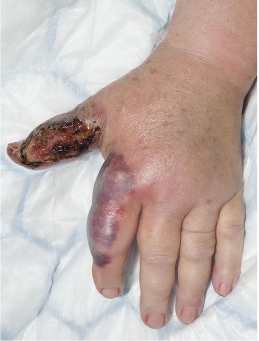
The plaques grew and the skin deteriorated with intravenous antimicrobials alone.
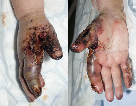
The debridement had no effect and the bullae grew, along with new lesions on non-adjacent digits.
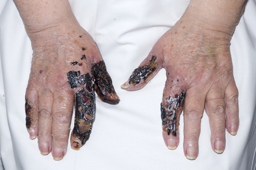
The hands at a clinical appointment at 1 month.
The cutaneous manifestations of the haematological malignancies are uncommon, but well documented [1]. They can be due to infiltration of tumour cells into the skin (leukaemia cutis), or due to a group of secondary reactions. The latter group includes Sweet syndrome, also called acute febrile neutrophilic dermatosis [2]. This is not a vasculitis, but behaves similarly with abrupt onset of tender purple papules on the upper extremities that are accompanied by fever and respond very well to steroid therapy. Sweet syndrome lesions are characterized by histology, with inflammatory changes and neutrophilic infiltrate [3]. The main differential diagnoses considered in this case were pyoderma gangrenosum, paraneoplastic pemphigus and the vasculitides, all of which can present as ulcerative lesions. Sweet syndrome was the primary working diagnosis due to the abrupt onset of the plaques, the histopathology, associated fever, raised CRP and excellent steroid response. Paraneoplastic pemphigus preferentially involves oral mucosa and typically responds poorly to steroid therapy [4]. Pyoderma gangrenosum can be associated with malignancy, but is more commonly seen in systemic inflammatory disorders, and usually affects the trunk and lower extremities. There was no evidence of vasculitis in the histology analysed.
The importance of all these lesions is that, as in this case, the skin is not improved by debridement. Immunosuppressive therapy should form the basis of management, and the vast majority of these lesions will respond to steroids.
CONFLICT OF INTEREST STATEMENT
None declared.
