


Abstract
The unique requirements of soft tissue coverage of the hand offer a challenge for the surgeon dealing with such defects. Local excision of such lesions may end in a defect that is not amenable to primary closure. Management of such defects requires the application of a graft or flap. We describe the application of a rotation flap for closure of a large defect on the dorsum of the hand following excision of a keratoacanthoma. The rotation flap offers repair with local skin of similar colour, texture and thickness. The end results were excellent both functionally and cosmetically.
INTRODUCTION
Skin lesions are common on the dorsum of the hand. Local excision of such lesions may end in a defect which is not amenable to primary closure [1]. The reconstructive ladder is based on the principle of using the simplest approach of coverage that adequately restores form and optimizes function. The ladder begins at direct primary closure, followed by skin grafting, local and regional flaps, and ends at free tissue transfer [2, 3].
A local flap, when available, offers the most convenient reconstruction and offers a repair of ‘like with like’ [1]. In this report, we describe the application of rotation flap to cover a large defect on the dorsum of the hand following excision of a keratoacanthoma.
CASE REPORT
A 94-year-old left-handed lady presented to our surgical clinic with a slowly growing lesion on the dorsum of the left hand, and the lesion had been increasing in size for the past few months interfering with the function of her dominant hand. In her background history, she had Type 2 diabetes.
On physical examination, there was a large dome-shaped lesion overlying the distal halves of the third to fifth metacarpals near the metacarpophalangeal joints (MCPJ) (Fig. 1).
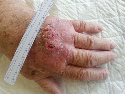
Clinical appearance of the keratoacanthoma located on the dorsum of the left hand.
The large size of the lesion, proximity to the MCPJ and being on the dominant hand of a highly independent patient represented extra challenge in the management. We decided to use a rotation flap to close the defect aiming at minimal functional impairment.
The procedure was carried on as an outpatient with local infiltration with 2% lignocaine with adrenaline.
The lesion was outlined, and the defect was triangulated with the apex pointing towards the dorsal wrist crease leaving a defect of 5 cm in diameter. Then, the flap was designed through a curved incision from the defect on the radial border (Fig. 2). The flap was raised using a scissors-spreading technique preserving the vessels and cutaneous nerves as much as possible.
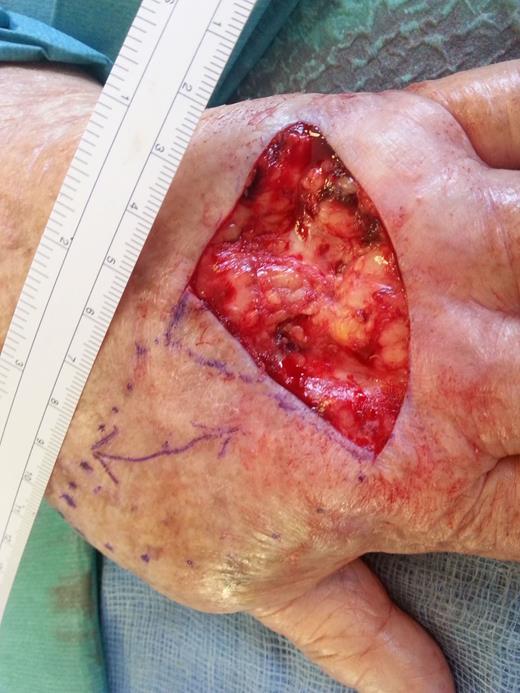
A triangulated defect following the local excision and the flap designed with a semicircular curve on the radial aspect.
Due to the large size of the defect, a back cut was done towards the apex of the primary defect to allow for extra rotation of the flap and less tension on the repair.
The flap then was rotated into the defect and sutured in place with 5/0 prolene stitches.
The secondary defect was closed by advancement using the laxity at the distal wrist crease (Fig. 3). The histology report showed a keratoacanthoma that was completely excised.
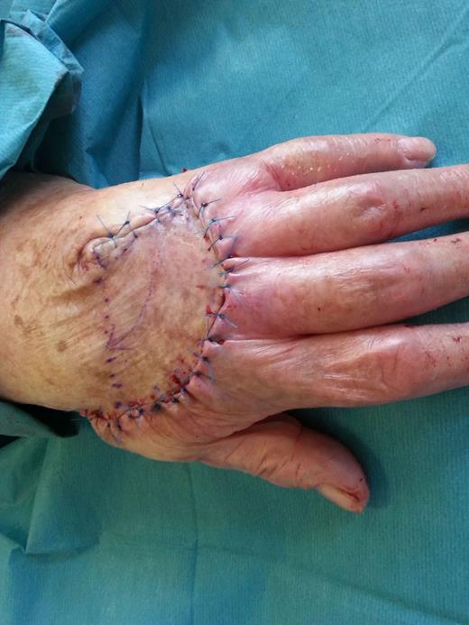
The flap rotated and sutured in position.
During the first visit to the clinic, 1 week later, the patient developed a superficial infection with some sloughing at the edges. This was satisfactorily managed with a course of antibiotic and repeated dressings; no revision of the flap was needed. Figure 4 shows the final outcome, 7 weeks post-operative, with excellent cosmetic results without functional compromise.
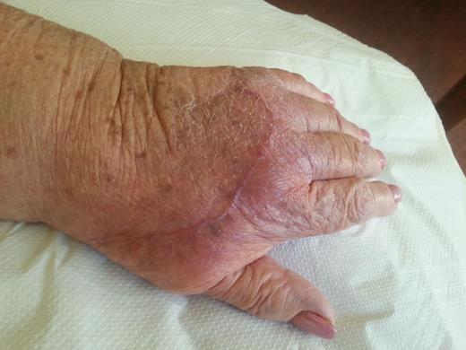
The end result of the flap in Figs 1–3.
DISCUSSION
The rotation flap is a well-established technique described in texts of plastic surgery dividing the tension of closure over a much larger surface area [3]. This technique has proved useful in reconstruction of defects up to 3 cm in diameter on the dorsum of the hand overlying the distal half of the metacarpals [1, 4]. In the present case, the defect was 5 cm in diameter on the dominant left hand.
The ideal soft tissue reconstruction should protect against the development of contractures and facilitate tendon and joint mobility, while maintaining durability and sensibility of the hand [2]. Split skin grafts give good results on the dorsum of the hand. However, the disadvantages include requiring a donor site, the quality of sensation is never good and the developing contracture. These make them not ideal in view of the functional requirements of the hand. The rotation flap, using a local tissue to replace a primary defect, adheres to the principle of replacing ‘like with like’ [1, 4].
The dorsal hand skin in elderly patients tends to be inelastic and does not lend itself to advancement. When a flap is used to reconstruct a defect, this creates a secondary defect. If the secondary defect is on the dorsum of the hand, it can no more be directly closed than the primary defect posing a similar problem. The principle of rotation flap on the dorsum of the hand is to use the laxity of skin at the dorsal wrist crease in the closure of the secondary defect [1].
It is recommended to apply the rotation flap to older patients with an element of actinic damage. The scar formed by such patients' skin on the dorsum of the hand tends to offer very good cosmetic and functional results. The use of similar extensive incisions in younger patients may not yield the same inconspicuous scars [1].
The rotation flap has an element of vascular axiality as the axially arranged venous plexus on the dorsum of the hand is preserved during raising the flap. This should at least in theory make the flap more robust and resistant to infection [1].
The excellent cosmetic results obtained with this technique are dependent on wound healing with minimal scarring. Dorsal hand skin in older patients, particularly with actinic damage, tends to heal with minimal scarring. Significant actinic damage would be a relative indication for this procedure in younger patients; otherwise, it is probably best reserved for patients older than 60 years [1].
In conclusion, we are reporting the application of the rotation flap principle to reconstruct a large defect on the dorsum of the hand. The flap offers repair with local skin of similar colour, texture and thickness. The end results were excellent both functionally and cosmetically.
CONFLICT OF INTEREST STATEMENT
None declared.
