


Abstract
Non-traumatic ureteral rupture has been reported more frequently, resulting from increased intraluminal pressures from distal urinary tract obstruction. We report the case of a 77-year-old man presenting with chronic urinary retention secondary to massive prostatic enlargement through acute kidney injury. Ultrasound scan detected a shallow left perinephric fluid collection with a possible bladder mass, demonstrated on flexible cystoscopy to be a massive median lobe of prostate. Computed tomography confirmed extravasation of urine from the left proximal ureter. In the absence of specific symptoms, the patient had successful conservative management with antibiotics and urinary catheterization for his acute episode, although declined further surgical intervention.
INTRODUCTION
Ureteral rupture is a urological emergency usually presenting with acute pain and requiring urgent surgical intervention. We report an unusual case where this condition was discovered incidentally on imaging, in the absence of any specific symptoms, to investigate chronic urinary retention caused by massive prostatic enlargement.
CASE REPORT
A 77-year-old Caucasian gentleman was admitted with acute kidney injury discovered on blood tests. He was known to have high-grade prostate intraepithelial neoplasia and atypical small acinar proliferation from prostate biopsies performed for a raised prostate-specific antigen (PSA), with a bone scan negative for potential metastases and was on PSA surveillance with the latest reading of 43 pre-admission. Prior to admission, he had lower urinary tract symptoms in the form of frequency, nocturia, intermittent urinary stream, terminal dribbling and a feeling of incomplete emptying. There was no significant past medical history. On examination, he was haemodynamically stable and afebrile. There was a palpable non-tender bladder with dullness to percussion and no renal angle tenderness.
Blood tests revealed a Stage 3 acute kidney injury (baseline renal function was normal) with raised inflammatory markers and elevated PSA compared with baseline (Table 1). A mid-stream urine specimen sent for microscopy, cultures and sensitivities yielded no growth of organisms. A bedside bladder scan revealed a bladder volume of >999 ml, necessitating the insertion of a urethral catheter with a residual volume of 2.4 l, and intravenous antibiotics were commenced. An ultrasound scan (USS) of the urinary tract was performed to evaluate for the presence of hydronephrosis; but while this demonstrated a minor dilatation of the left renal pelvis, it also found a shallow fluid collection around the left kidney extending into the left lateral retroperitoneum, separate from the left psoas muscle (Fig. 1). In the urinary bladder, there was an irregular 4 cm mass on the left posterolateral aspect. In view of the findings, an urgent flexible cystoscopy was performed, revealing a massive median lobe of the prostate protruding up and back into the bladder; the ureteric orifices were not visualized because of this, and the bladder mucosa was normal. The patient subsequently underwent a computed tomography (CT) urogram, which showed bilateral fullness in the pelvicalyceal systems and confirmed a 4.8 × 4.4 × 2.8 cm fluid collection adjacent to the lower pole of the left kidney as detected on the USS, with perinephric stranding. The fluid collection was demonstrated to be extravasated urine on the delayed post-contrast images arising from a defect in the left proximal ureter, and extending down the left paracolic gutter (Figs 2 and 3). The massive prostate was also demonstrable (Fig. 3).
Blood results on admission
| Haematology | |
| Haemoglobin | 101 g/l |
| White cell count | 11.7 × 10⁹/l |
| Platelet count | 204 × 109/l |
| Biochemistry | |
| Sodium | 139 mmol/l |
| Potassium | 4.6 mmol/l |
| Urea | 38.7 mmol/l |
| Creatinine | 305 mmol/l |
| Estimate glomerular filtration rate | 17 ml/min/1.73 m2 |
| Prostate-specific antigen | 71.8 ng/ml |
| C-Reactive protein | 295 mg/l |
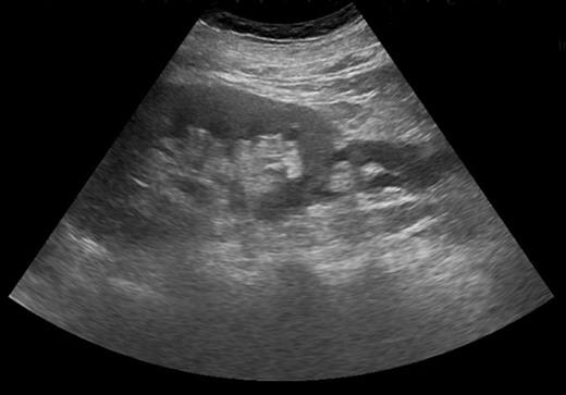
USS demonstrating shallow fluid collection around left kidney.
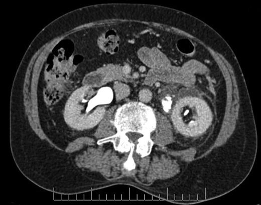
Axial section CT image demonstrating defect in left proximal ureter from which contrast is extravasating, with perinephric fluid collection and fat stranding.
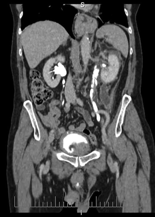
Coronal section CT image demonstrating fluid collection extending down left paracolic gutter. Also visible are massively enlarged prostate and urinary extravasation from left ureter.
Management options were discussed with the patient in the form of conservative treatment with antibiotics and catheterization, or insertion of a nephrostomy as retrograde ureteral stenting would have not been possible due to prostatic size. He opted for the conservative approach which was successful with resolution of renal function and inflammatory markers to normal parameters. He was offered a Millins' prostatectomy but declined and was happy to be managed with a long-term catheter and repeat PSA measurement in 3 months' time.
DISCUSSION
Non-traumatic ureteral rupture has been reported more frequently in the recent literature despite its unusual occurrence. Physical obstruction distally within the urinary tract, most commonly by a ureteral calculus, is thought to transmit increased intraluminal pressures proximally into the ureter [1]. Other reported causes of such obstruction include gravid uterus, urinary tract malignancy and even iatrogenic surgical ligation of the ureter [2–4]. Urinary retention has previously been described as a cause of ureteral rupture, but this was in the presence of abdominal pain [5], reported also in other cases when urine has leaked either into the retroperitoneal space or the peritoneal cavity. The absence of specific abdominal symptoms in this particular case was unusual and therefore meant that the incidental discovery of a ruptured ureter on imaging was unexpected, albeit probably not surprising given the prostatic size.
Severe intractable abdominal pain similar to that attributed to renal colic is the most observed presentation of ureteral rupture [1, 6], and its presence is confirmed on imaging, which may be performed to investigate other causes of pain. CT scan of the urinary tract with a delayed excretory phase of contrast is likely to be the most used means and can determine the presence of extravasation, likely location of rupture and extent of urinoma or abscess formation, as well as the nature of the obstructing lesion. If retrograde ureteral stent placement is to be performed, then pyelography can be performed at the same time to confirm CT findings. Ultrasonography has a limited role for detecting ureteral pathologies but was key here in detecting a perinephric fluid collection and possible bladder mass, which led to further investigations.
Many reported cases of non-traumatic ureteral rupture have involved acute surgical management in the form of percutaneous drainage of urinomas or fluid collections, and urinary diversion via insertion of percutaneous nephrostomies and placement of ureteral stents [2, 4, 6]. In our case, however, factors that obviated this were the absence of any clinical signs or symptoms of ureteral rupture, the patient's improving clinical condition and the massive median lobe of prostate preventing visualization of the ureteric orifices at flexible cystoscopy and thus making potential retrograde ureteral stent insertion difficult and complicated. There is documented evidence of successful conservative treatment with subsequent reabsorption of urine [7], and our case is an additional demonstration of this.
This case also highlights what is now documented to be a rare but serious complication of high-pressure chronic urinary retention. Although there is a previous report of ureteral rupture caused by neurogenic urinary retention [5], to our knowledge, this is the first case in a male patient with bladder outflow obstruction from prostatic enlargement. In a situation of chronic retention where high pressures are suspected present as indicated by acute kidney injury, when performing an USS one must be open to the possibility of ureteral rupture occurring even in the absence of symptoms.
CONFLICT OF INTEREST STATEMENT
None declared.
